


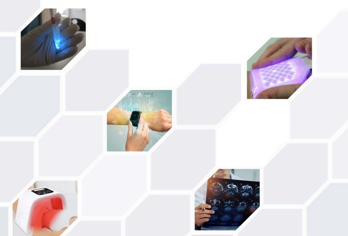
The use of light in medicine dates back to ancient civilizations, when sunlight was employed to treat various ailments. Today, with advancements in technology, the medical community has harnessed specific wavelengths of light to develop targeted treatments for a wide range of conditions.
From ultraviolet (UV) to visible (VL) and infrared (IR) light, each wavelength interacts uniquely with human tissues, offering therapeutic benefits and presenting potential risks. Understanding these interactions is important for optimizing the use of light in medical applications while ensuring patient safety.
Understanding how various wavelengths penetrate different layers of skin and tissue is key to maximizing therapeutic benefits while minimizing risks. Each wavelength interacts uniquely with human tissue, providing specific advantages and challenges in medical applications.
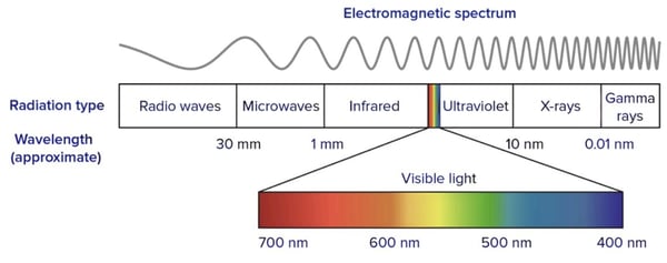
Source: Wavelengths of light and photosynthetic pigments
Ultraviolet (UV) Light
UV light, ranging from 100–400 nanometers (nm), is divided into three categories¹:
UV-A (320–400 nm): Penetrates the dermis; used in phototherapy for conditions like psoriasis and vitiligo. Prolonged exposure can cause skin aging and increase cancer risk.
UV-B (290–320 nm): Penetrates the epidermis; effective in treating skin conditions by slowing cell growth. Also administered in controlled doses to prevent sunburn and lower cancer risk.
UV-C (100–290 nm): Primarily used for sterilization due to its germicidal properties. Science has shown 208-210 nm range has strong germicidal effects, able to inactivate bacteria and viruses on skin without harming human tissue.
Visible Light (VL)
Visible light takes up the 400–700 nm spectrum and is perceived as different colors, each offering unique therapeutic benefits¹:
Blue light (440–500 nm): Penetrates the upper dermis; effective for neonatal jaundice and acne. Antibacterial properties also help eliminate bacteria on the skin's surface.
Green light (500–570 nm): Penetrates the upper dermis and is used to treat pigmentation issues by targeting melanocytes.
Yellow light (570–590 nm): Penetrates the dermis and is known for its ability to stimulate collagen production, improve skin texture and also has a calming effect on the nervous system.
Red light (620–750 nm): Penetrates the dermis and subcutaneous tissue, known for antiinflammatory and wound-healing properties.
“The various wavelengths of light are harnessed in wideranging medical applications that leverage their unique properties to treat numerous conditions.”
Infrared (IR) Light
Infrared light ranges from 700 nm to 1 mm and is categorized into three types¹:
Near-Infrared (NIR) light (700–1.400 nm): Penetrates muscles, nerves, and bones, and can be effective for pain management and deep tissue repair.
Mid-Infrared (MIR) light (1,400–3,000 nm): Provides thermal benefits; used in therapies requiring moderate heat.
Far-Infrared (FIR) light (3,000 nm–1 mm): Penetrates surface layers of the skin, providing gentle heating beneficial for circulation and reducing stiffness.
Various wavelengths of light are harnessed in different medical applications that leverage their unique properties to treat numerous conditions.
Optical Biosensors
Optical biosensors are advanced analytical devices that use light to detect and analyze biological molecules and/or chemical substances. These sensors measure changes in light properties, such as absorbance, fluorescence, luminescence, or reflectance, caused by the interaction between a target analyte and a biorecognition element on the sensor surface.
Optical biosensors are highly valued in healthcare, environmental monitoring, and biotech for their high sensitivity, specificity, and real-time analysis capabilities. Factors such as color temperature, which affects fluorescence excitation and signal stability, can influence their performance, making them crucial tools for noninvasive, multifaceted, and continuous health monitoring⁴.
Photobiomodulation (PBM) for Wound Care
Light therapy is emerging as a promising technique for accelerating wound healing, particularly for severe and chronic wounds that are challenging to treat with conventional methods. Light therapy uses specific wavelengths of light to interact with tissues and cells, promoting natural healing processes by accelerating mitochondrial production of ATP and thereby reducing inflammation, minimizing scarring, and accelerating tissue repair.
Unlike traditional methods, light therapy is minimally invasive and often provides faster healing times. Wound care light therapy applications include treating burns, surgical wounds, and even non-healing wounds, making it a valuable tool in modern wound care.⁵
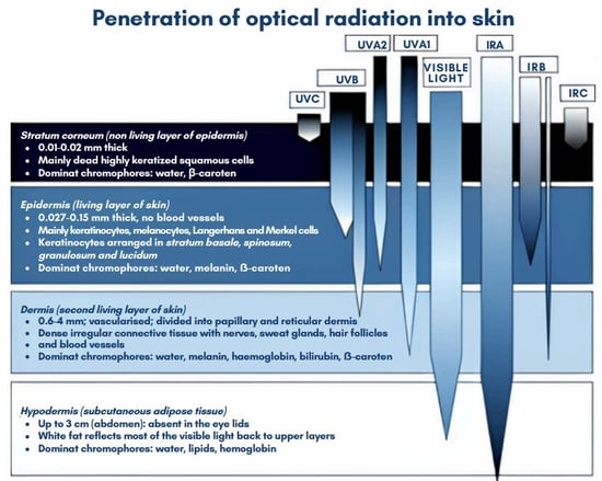
Source: Optical radiation in modern medicine (National Library of Medicine; 2013)
Photobiomodulation in Neurological Applications
PBM therapy is emerging as a catalytic, non-invasive treatment for various neurological disorders, including Alzheimer's, Parkinson's, and different traumatic brain injuries.
This innovative therapy involves the use of low-intensity red or near-infrared (NIR) light to stimulate neural activity and enhance brain function. By targeting cytochrome c oxidase in mitochondria, PBM therapy promotes cellular survival, reduces apoptosis, alleviates oxidative stress, and suppresses inflammation, potentially leading to significant improvements in cognitive and motor functions.
Clinical studies have shown PBM's potential to improve memory, attention, and mood in Alzheimer's patients, reduce motor impairment in Parkinson's patients, and enhance cognitive functions in TBI patients. As research progresses, PBM therapy may offer new hope for individuals suffering from debilitating neurological conditions.⁶
“As research progresses, PBM therapy continues to offer new hope for individuals suffering from debilitating neurological conditions.”
Bioluminescence in Medicine
Bioluminescence, the natural phenomenon by which living organisms emit light, has recently gained traction in medical research due to its potential in diagnostics, imaging, and therapeutic techniques.
Most types of bioluminescence involve a chemical reaction between luciferin, a lightemitting molecule, and luciferase, an enzyme, which produces light efficiently without significant heat generation.⁹
Observed by figures of antiquity like Aristotle and Pliny the Elder, bioluminescence has transitioned from a curiosity of the natural world to a tool with powerful potential in modern medicine. Bioluminescent markers enable researchers to visualize and track diseases by incorporating luciferase genes into cells, which emit light when a substrate like luciferin is present. This non-invasive imaging technique allows for real-time monitoring of disease progression and treatment efficacy in live animal models, providing critical insights into cancer growth, metastasis, and infectious disease dynamics.⁹
Additionally, bioluminescence facilitates drug discovery and development, enabling rapid and sensitive screening of potential therapeutic agents. As advancements continue, bioluminescence could potentially transform non-invasive diagnostics.
While light therapy offers numerous benefits, it’s not always without risks. Understanding these risks is critical for safe light therapy applications in medical treatments.
UV Radiation Risks
UV radiation can cause acute skin damage, manifesting as erythema similar to sunburn. Chronic exposure may lead to photo-aging and a higher risk of skin cancers like melanoma.
The eyes are particularly sensitive to UV radiation. Too much UV light could lead to conditions like photokeratitis and conjunctivitis, which can also contribute to cataract formation.⁷
Visible Light Risks
Blue light, though beneficial for specific medical treatments, can cause retinal damage with prolonged exposure, requiring regulated exposure to prevent eye strain and potential macular degeneration. Additonally, some people have a nausea reaction to blue light.⁷
Infrared Radiation Risks
The primary concern with infrared radiation is thermal damage, including skin burns and heat-induced rashes. Chronic exposure might lead to conditions like erythema ab igne (EAI), increasing the risk of skin cancer. However, these risks are generally associated with improper use or excessive exposure.⁷
The powerful potential of light in medicine is to offer non-invasive diagnostics, realtime disease monitoring, and targeted treatments.
While understanding and mitigating associated risks is crucial for patient safety, the integration of light-based technologies promises improved patient outcomes, reduced treatment times, and lower healthcare costs, promising a brighter future for medical advancements.
"The powerful potential of light in medicine is to offer non-invasive diagnostics, realtime disease monitoring, and targeted treatments."

Optical biosensors are cutting-edge medical devices that use light to detect and analyze biochemical substances and reactions. They track changes in optical properties like absorbance and fluorescence, enabling noninvasive, highly sensitive, continuous health monitoring.
Key advantages of optical biosensors include their compact size, ability to perform multiple tests on a single sample, and seamless integration with wearable devices like smartwatches.
This article explores the fundamentals of optical biosensors and their numerous applications and advantages. It highlights recent innovations, such as silicon photonics that are driving advancements in this field.
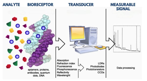
Optical biosensors provide significant advantages in both medical and life science applications, making them ideal for diagnostic wearable devices, continuous health monitoring, and patient comfort.
Non-Invasive Detection
The most obvious advantage of optical biosensors is their non-invasive nature. They don’t always require direct contact with samples, making them ideal for continuous monitoring of various physiological parameters.
This is why optical biosensors are so popular in wearable devices, where sensors can easily and discreetly monitor health metrics without the patient noticing.
“Immediate, 24/7 feedback and timely medical interventions allow patients to remain at home vs. a care facility and enhances patient care and treatment outcomes.”
If you’ve ever looked at the underside of your Fitbit, for example, you might have noticed the blinking green light—that’s an optical biosensor. This green LED light is used to measure your heart rate. It works by shining light into your skin and measuring the amount of light that is reflected back.
Blood absorbs green light, so the sensor can detect changes in blood flow by analyzing the variations in the reflected light. This method allows the device to continuously monitor your heart rate without any discomfort.
Multiplexing Capability
Optical biosensors can analyze multiple analytes simultaneously from one sample, such as a single blood draw. This ‘multiplexing’ capability enhances their efficiency in clinical diagnostics and research, enabling comprehensive analyses with minimal invasiveness.
Compact Size
The gradual miniaturization of optical biosensors allows for the development of more portable and discreet wearable devices. These compact sensors are well suited for continuous health monitoring, providing real-time data even while the patient is sleeping.
Real-Time Analysis
Optical biosensors offer noninvasive real-time analysis for monitoring dynamic biological processes. This allows for immediate, 24/7 feedback and timely medical interventions, allowing patients to remain at home vs. a care facility, and enhancing patient care and treatment outcomes.
Wearability
The integration of optical biosensors into wearables has significantly advanced personal health monitoring. It allows for real-time data collection and analysis, which can be synced with mobile apps and other digital health platforms. This seamless integration helps users make informed decisions about their health and lifestyle, enhancing overall well-being and potentially preventing serious health issues.
Optical biosensors are versatile tools with a wide range of applications in medical and health monitoring.
Glucose Monitoring
Current healthcare expenditures on diabetes management are significant.⁵ Wearable optical biosensors, such as colorimetric sensors in contact lenses, offer non-invasive and continuous glucose monitoring. By reducing the reliance on costly glucose monitoring, these innovations have the potential to lower healthcare costs. Coupled with advancements in light therapy, these technologies could not only improve patient outcomes but also address the urgent need to decrease costs on glucose monitoring.⁵
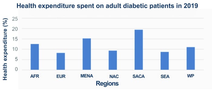
Source: Optical sensors for continuous glucose monitoring (IOPScience; 2021)
Vital Signs Monitoring
Optical biosensors are widely used in wearables to monitor heart rate, respiration rate, blood pressure, and more. These sensors provide real-time data, aiding in the early detection and management of cardiovascular conditions.⁶
Electrolyte and pH Monitoring
Colorimetric optical biosensors can measure electrolyte concentrations and pH levels in bodily fluids. These sensors are essential for maintaining electrolyte balance and monitoring conditions like acidosis or alkalosis.⁷
Optical biosensors and electrical sensors differ primarily in how they detect signals. Electrical sensors measure changes in electrical properties like resistance or current, while optical biosensors detect changes in light properties such as fluorescence or absorbance.
One of the main benefits of optical biosensors is their high specificity. They can detect specific molecules with great precision, even in complex biological environments. This is important for sensitive applications, like medical diagnostics. Additionally, optical biosensors provide real-time analyses, offering immediate feedback for timely interventions.
Optical biosensors also excel in environments where electrical interference might affect performance. Electrical sensors can be impacted by the presence of other ionic species or electrical noise, leading to inaccurate results. Optical sensors using light-based methods are less likely to be affected, making them more reliable in complex settings like in vivo monitoring or environmental testing.⁶
“Wearable technologies use light to measure changes in blood flow or oxygen saturation, offering a noninvasive way to track vital signs throughout the day.”
Optical and electrical sensors can also be used together. Combining both technologies can provide comprehensive data, improving accuracy and reliability. For instance, while an optical sensor might offer precise biomolecule detection, an electrical sensor can measure related electrical changes, providing a multidimensional view of the analyte in question.
Watch devices incorporate optical biosensors to monitor various health metrics, including heart rate and blood oxygen levels. These wearable technologies use light to measure changes in blood flow or oxygen saturation, offering a noninvasive way to track vital signs throughout the day.⁴
Some advanced wearables are capable of monitoring glucose levels or a person’s hydration. These evolving devices use sophisticated optical biosensors that can detect a slight change in skin and interstitial fluid, providing valuable, real-time data for managing conditions like diabetes while ensuring adequate hydration.⁷ The accuracy and reliability of these devices have improved with advancements in optical techniques, which measure skin hydration by analyzing light penetration.
These methods assess the water content in the skin's outermost layer, crucial for maintaining skin barrier function. By monitoring these parameters, these devices can provide a reliable tool for various applications.⁷
Innovative applications of optical biosensors include ‘tattoo-based’ sensors for monitoring pH, glucose, and sodium levels. These tattoos change color in response to the concentration of specific analytes, providing a visual and non-invasive method of monitoring health parameters.
Silicon photonics integrates sensor technology with existing semiconductor manufacturing, leveraging established infrastructure to produce optical sensors on a large scale.
This approach means it is now possible to develop compact, portable biosensors that are even better suited for discreet wearable devices and real-time diagnostics while maintaining high sensitivity and specificity.
Additionally, silicon photonics manufacturing processes significantly lower production costs, promoting broader adoption and enhancing public health monitoring capabilities. Silicon photonics also improves sensor integration with other electronic components, enhancing functionality and reliability.⁴
Optical biosensors represent a cutting-edge technology with significant potential in medical diagnostics, personal health monitoring, and many other applications. Their non-invasive nature, real-time analysis capabilities, and adaptability make them very useful tools in modern healthcare, particularly for remote health monitoring devices.
As advancements in LED, Laser Diode, and OLED technologies continue to evolve, optical biosensors can become even more cost effective, efficient and versatile. Laser diodes, for instance, offer precise light sources used in smartwatches for continuous health monitoring. The evolution of these lighting technologies directly enhances the accuracy of optical biosensors, enabling more precise analysis. Looking ahead, the integration of these lighting solutions with optical biosensors will drive future innovations in healthcare.
"Optical biosensors represent a cutting-edge technology with significant potential inmedical diagnostics, personal health monitoring, and many other applications."


Wound healing is a complex process that can be hindered by various factors such as diabetes, obesity, hypoxia, and infections. Traditional treatments often fall short, leading to prolonged recovery times and increased risk of complications.
Light therapy, using LED and laser light sources, has emerged as a promising technique to accelerate wound healing. This is especially true for severe wounds such as burns, surgical wounds, and chronic non-healing wounds, which are difficult to treat with conventional methods.
Unlike negative pressure wound therapy, which can be invasive and induce inflammation, light therapy is minimally invasive and often provides quicker healing times. Additionally, light therapy can be more eco-friendly, using reusable devices and reducing the need for disposable materials.
“Light therapy, using LED and laser light sources, has emerged as a promising technique to accelerate wound healing.”
Studies have shown that light therapy can significantly reduce pain, accelerate healing times, minimize scarring, and reduce inflammation. It employs different wavelengths of light, each interacting with the body in unique ways to promote healing.
Wavelength and Spectrum Various wavelengths of light play distinct roles in the wound-healing process, each interacting with tissues and cells in unique ways.
Violet and Blue light (380–500nm) are primarily known for their potent antibacterial properties, which can sterilize wounds without hindering the healing process. By targeting bacterial cell walls and disrupting their structure, blue light therapy can help prevent infections that commonly impede wound healing. These wavelengths can penetrate the skin to reach superficial wounds and are often used to treat infected, non-healing wounds.⁸
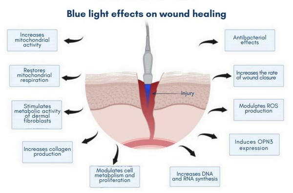
Red (620–750nm) and Near-Infrared (750–950nm) wavelengths penetrate deeper into the tissue compared to violet and blue light. These wavelengths are highly effective at encouraging tissue repair and regeneration by interacting with chromophores like cytochrome c oxidase in mitochondria. This interaction enhances cellular respiration and energy production, which accelerates the healing process.
Red and near-infrared light also stimulate collagen production, reduce inflammation, and improve blood circulation to affected areas. These benefits make red and nearinfrared light therapy ideal for treating deeper tissue wounds, such as ulcers and burns, where enhanced tissue repair and reduced inflammation are crucial for recovery.⁴
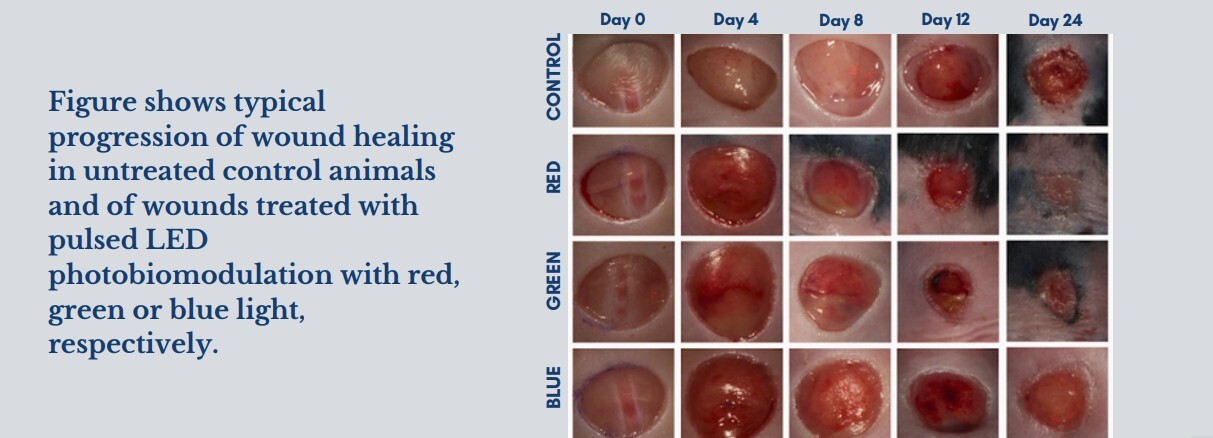
The intensity and exposure duration of light therapy are critical factors in determining its effectiveness for wound healing. Patients typically undergo 6 to 12 sessions, with each session lasting from a few minutes to 30 minutes, depending on the severity and type of wound.
The optimal energy density for effective treatment is dependent on the depth of the wound and the wavelengths utilized. Studies have shown that there is a minimum energy density levels in the range from 5.0 – 40 J/cm². Treatment effectiveness is dependent on both the energy density and the treatment duration. It is critical to optimize these factors to stimulate cellular activity and promote healing without causing damage. Treatments with energy densities above 50 J/cm² can be less effective or even harmful, potentially leading to cell damage or death.
“Modern light therapy treatments integrate certain wavelengths with other features to optimize healing outcomes.
Studies have shown that using blue and near UV light inhibits bacterial growth and can be used initially to minimize the bacterial load. Treatment can be followed with red light to enhance tissue repair and closure. The specific treatment protocol can also vary depending on the type of wound and the patient’s overall health.
For instance, chronic wounds with a high bacterial load might benefit from more frequent blue light sessions to inhibit bacterial growth. The exposure time and fluence levels for this to work would need to be very high and prepare the wound for subsequent healing phases. On the other hand, red and near-infrared light therapy might be more beneficial during the later stages of wound healing, focusing on reducing inflammation and promoting tissue regeneration.
Modern light therapy treatments integrate certain wavelengths with other features to optimize healing outcomes.
For instance, hydrofiber silver integrates soft non-woven fibers with ionic silver, providing antimicrobial properties while maintaining a moist wound environment. When used in tandem with blue light therapy, hydrofiber silver dressings can effectively reduce bacterial load, leveraging the antimicrobial action of both the light and the silver ions. This dual approach ensures a sterile environment conducive to faster healing.
Another example is hydrogel, a gel-based solution designed for optimal wound healing. Red and near-infrared light therapy can be used with hydrogel to enhance collagen production and tissue repair.
The hydrogel keeps the wound moist and provides a cooling effect, accelerating the healing process and minimizing scarring when combined with light therapy.⁵
Patient comfort is a top priority in the design of light therapy devices. Many of these devices are ergonomically designed to ensure they can be used comfortably over the treatment area. Adjustable intensity settings allow for customization based on the patient's pain threshold and specific wound requirements, reducing the risk of discomfort during therapy.
Moreover, sessions can be brief, which minimizes disruption to the patient’s daily routine. The non-invasive nature of light therapy, coupled with the thoughtful design of these devices, enables a pleasant treatment experience, encouraging consistent use and adherence to prescribed therapy regimens.
Several devices are currently available for wound healing using light therapy:
One such device uses red light to accelerate wound healing by promoting increased blood flow and collagen production. It is portable and user-friendly, making it ideal for both clinical and home use.
Designed for professional use, a medical-grade red light therapy pad has been created to deliver concentrated red light therapy to larger wound areas, enhancing cellular repair and reducing inflammation.
A Photobiomodulation device has been developed to offer both red and nearinfrared light therapy, providing comprehensive treatment for various wound types. It can be adjusted to cater to different treatment needs.
Combining electrotherapy with light therapy, there is a system that offers a multifaceted approach to wound healing, particularly effective for chronic and difficult-to-heal wounds.
Another product on the market is an advanced laser system that provides targeted light therapy for wound healing, reducing pain, and accelerating tissue repair. It’s suitable for clinical settings, offering precise and effective treatment options.
Advancements in light therapy enable an important step forward in wound care. This innovative approach offers a minimally-invasive and effective treatment option, and also aligns with eco-friendly practices by reducing the need for disposable materials.
As technology and research continue to progress, the potential of light therapy is likely to expand, offering more refined and effective treatments. Healthcare professionals have the opportunity to embrace this catalytic technology, enhancing patient care and setting a new standard in wound management.
“The non-invasive nature of light therapy, coupled with thethoughtful design of these devices, enables a pleasant treatment experience, encouraging consistent use and adherence to prescribed therapy regimens.”


Neurological disorders affect millions of people worldwide, causing profound personal and societal injury. Conditions such as Alzheimer's, Parkinson's, and traumatic brain injury (TBI) devastate individual lives and strain healthcare systems.
The quest for effective, non-invasive treatments has led to exploration of innovative modalities, including light therapy. Emerging research such as Dr. Michael Hamblin's pioneering work in his book 'Photobiomodulation in Neuroscience,' highlights the potential of light as a powerful healing tool, offering new hope for treating neurological disorders.
Brain photobiomodulation (PBM) therapy stands out as a promising noninvasive treatment modality. It harnesses the healing power of light to stimulate neural activity and improve brain function.
Brain photobiomodulation (PBM) therapy is an innovative treatment that uses lowintensity light to stimulate neural activity and improve brain function.
This therapy involves exposing neural tissue to red or near-infrared (NIR) light, typically delivered through various methods such as LEDs or lasers.
The wavelengths of light used in PBM therapies range from 600–1,100 nm, with red light (up to 700 nm) and NIR light (up to 940 nm) primarily targeting cytochrome c oxidase in mitochondria. Longer NIR wavelengths (980 nm and 1,064 nm) affect heat-sensitive ion channels.
Molecular Mechanisms of PBM Therapy
Therapeutic effects of PBM start when light passes through the scalp and skull before it’s absorbed by specific molecules in the brain. Depending on the wavelength, the light interacts with two main chromophores: cytochrome c oxidase and transient receptor potential (TRP) ion channels.
For red and near-infrared (NIR) light up to 940 nm, the primary chromophore is cytochrome c oxidase, an enzyme in the mitochondrial respiratory chain of cortical neurons. When cytochrome c oxidase absorbs light, it increases the production of cellular energy (ATP) and reactive oxygen species (ROS).
These changes enhance cell signaling and overall mitochondrial activity, leading to improved cellular survival, reduced apoptosis, decreased oxidative stress, and suppressed inflammation. Collectively, these effects can enhance brain function and cognitive ability.
Longer wavelength NIR light (980 nm and 1,064 nm) is primarily absorbed by heat and light-sensitive TRP ion channels. These channels, particularly the transient receptor potential vanilloid (TRPV) type, respond to light by altering cellular ion flows and further promoting cell signaling and messenger molecule production.
Similar to the effects on cytochrome c oxidase, this stimulation of TRP channels leads to increased mitochondrial activity, boosting ATP and ROS production.
Photobiomodulation underlying mechanisms at the cellular and molecular levels
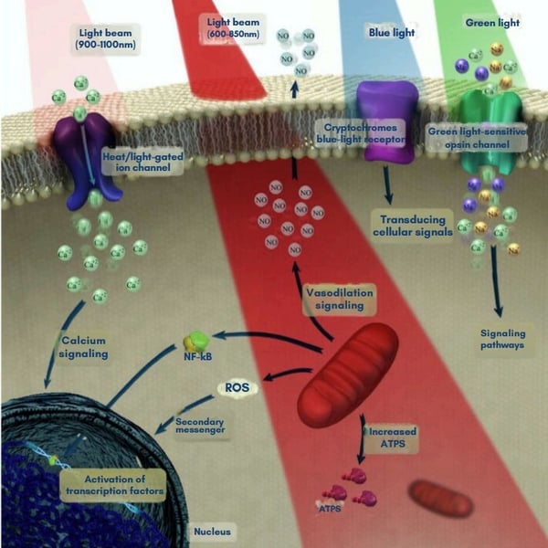
Source: Brain Photobiomodulation Therapy: A Narrative Review (National Library of Medicine; 2018)
Different approaches for light delivery to achieve brain photobiomodulation therapy
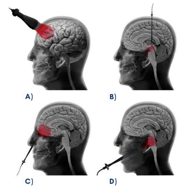
Source: Brain Photobiomodulation Therapy: A Narrative Review (National Library of Medicine; 2018)
Heat and Risk Factors
The use of PBM therapy involves managing potential heat risks. Light intensity needs to be carefully controlled to prevent overheating skin.
Research suggests that power densities for light therapy devices used on the head are typically kept below certain thresholds to avoid potential discomfort or adverse effects. Studies indicate that perceptible heating of the skin may begin when power density exceeds approximately 500 mW/cm². At higher levels, around 1 W/cm², the sensation of heat can become more pronounced.²
Proper device design is essential to ensure patient comfort and safety. Features that regulate temperature and distribute light evenly to prevent hotspots are best practice. By maintaining strict safety standards, PBM devices can effectively deliver therapeutic benefits without causing adverse effects.
Light Therapy Considerations
Effective PBM therapy also requires careful consideration of light wavelength, intensity, and exposure time. Wavelengths between 650–1,200 nm are most effective for treating neurological conditions. The intensity of light also matters: low doses might not have a significant effect, while high doses can actually harm cells.
It's important to find the right balance. Studies show that light needs to be delivered in specific doses to stimulate beneficial effects. For example, an optimal dose can boost cellular energy and improve mitochondrial function, but too much light can damage mitochondria. Proper device design also helps ensure that light therapies are delivered in the right dosage to maximize benefits and minimize risks.
Potential Outcomes and Ideal Candidates
The therapeutic outcomes of PBM are extensive and include improved cell survival, reduced apoptosis, decreased oxidative stress, and suppressed inflammation. A study by Salgado et al. showed significant improvement in cerebral blood flow in elderly women following transcranial LED therapy. These improvements were measured using transcranial Doppler ultrasound, indicating enhanced blood flow velocity and reduced resistance in key cerebral arteries.
For conditions associated with cognitive decline, like Huntington’s disease and diabetic retinopathy, PBM has demonstrated neuroprotective effects, highlighting its potential as a broad-spectrum therapy for various neurological disorders.
Brain tissue-specific functional processes that occur after brain photobiomodulation therapy
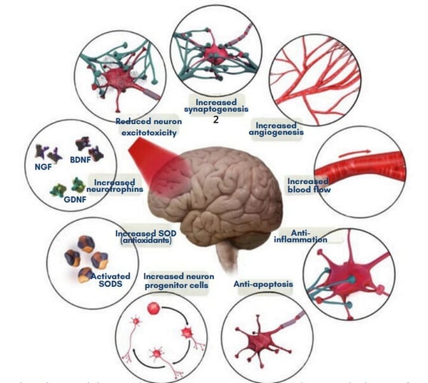
Source: Brain Photobiomodulation Therapy: A Narrative Review (National Library of Medicine; 2018)
Traumatic Brain Injury (TBI)
PBM therapy has been shown to improve outcomes for TBI patients, including enhanced self-awareness, social functioning, and sleep quality.
Transcranial LED therapy (633/870 nm) can also improve self-regulation in social functioning and sleep quality. Higher doses of NIR light have been particularly effective in reducing headaches and improving mood and cognitive states.
“PBM therapy has been shown to improve outcomes for TBI patients, including enhanced self-awareness, social functioning, and sleep quality.”
Another study investigated the effects of PBM on cerebral blood flow and cognitive function in patients with chronic TBI. The results showed significant improvements in executive function, memory, and sleep quality, suggesting that PBM can effectively enhance cognitive performance and overall brain health in TBI patients. In animal models, PBM has also been shown to reduce inflammation and cell death while improving cognitive functions such as learning and memory.
Alzheimer’s Disease
Research suggests that NIR PBM therapymay have positive effects on patients with Alzheimer's disease. Some studies have reported improvements in sleep duration, mood, and various aspects of cognitive function, including memory and attention, following NIR PBM treatments.⁶ Studies have demonstrated that PBM can reduce amyloid-beta plaques and tau protein tangles, which are hallmarks of Alzheimer’s disease, potentially slowing disease progression and improving patient outcomes.
“PBM therapy enhances cerebral blood flow and oxygenation, providing critical support to brain regions affected by Alzheimer’s.”
Additionally, PBM therapy enhances cerebral blood flow and oxygenation, providing critical support to brain regions affected by Alzheimer’s.
By improving mitochondrial function and reducing oxidative stress and inflammation, PBM helps protect neurons from damage and promotes neurogenesis, the creation of new neurons. This can contribute to better cognitive performance and could offer a non-invasive, low-risk treatment option to help manage and mitigate the symptoms of Alzheimer’s disease, improving patient quality of life.
Parkinson’s Disease
Research has shown that PBM therapy may be beneficial for Parkinson’s disease. studies have reported improvements in both motor and cognitive functions following PBM treatment.⁵ These improvements are attributed to the therapy’s ability to protect neuronal cells and stimulate neurogenesis.
In animal models of Parkinson’s disease, PBM has been shown to improve motor performance and reduce neuroinflammation. Clinical studies have reported improvements in motor skills and cognitive functions, suggesting that PBM could be a valuable adjunctive treatment for Parkinson’s patients.
While photobiomodulation devices for general health are widely available, the number of specific devices for transcranial applications is growing. These devices, often helmets or headbands with integrated NIR LEDs, are designed to target specific brain regions, providing non-invasive treatment options for neurological disorders.
There are specialized photobiomodulation (PBM) devices on the market designed for brain therapy, offering targeted treatment for conditions such as Alzheimer’s and traumatic brain injury (TBI).
Emerging research underscores the potential of PBM to enhance cognitive performance, reduce symptoms, and improve overall brain health.
By leveraging the therapeutic effects of light to stimulate neural activity, PBM therapy provides a safe and effective treatment option. As the understanding and application of this innovative therapy continue to grow, PBM holds great promise for transforming the management of neurological disorders and significantly improving patient outcomes and quality of life.
“By leveraging the therapeutic effects of light to stimulate neural activity, PBM therapyprovides a safe and effective treatment option.”


Bioluminescence, the natural emission of light by living organisms, has captivated human curiosity for centuries. From the mesmerizing glow of fireflies to the radiant depths of the ocean, bioluminescence is a fascinating phenomenon.
Recently, this natural wonder has found groundbreaking potential in the field of medicine, improving how various diseases are diagnosed, monitored, and potentially even treated. This article delves into the history, mechanisms, and medical applications of bioluminescence, highlighting its potential to enhance healthcare.
Bioluminescence is the production and emission of light by living organisms through a chemical reaction involving luciferin, a light-emitting molecule, and luciferase, an enzyme. Luciferase catalyzes the oxidation of luciferin, resulting in an excited state that emits light as it returns to its ground state.
This process is observed in many organisms, including over 75% of deep-sea creatures and common insects like fireflies. Currently, nine different luciferins have been identified among the 40 known bioluminescent systems, each capable of emitting different wavelengths of light, creating a variety of glow colors. This process is highly efficient, producing light without significant heat, making it an intriguing subject for scientific research and practical applications.
Bioluminescence reaction of Fluc, Rluc and Gluc.
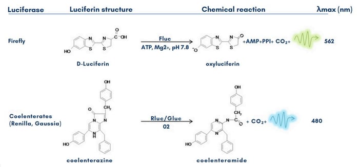
Source: Bioluminescence Imaging: Progress and Applications (NIH; 2011)
In the 21st century, bioluminescent applications are primarily confined to laboratory research involving animal models, such as rats, mice, and rhesus macaques.
These animal models enable researchers to study disease processes and test new treatments in a controlled environment. In vitro cell cultures are also used to investigate cellular responses to various stimuli, providing valuable data for preclinical studies.
While bioluminescence has yet to be widely applied in human medicine, its potential for non-invasive diagnostics and real-time monitoring holds promise for future clinical use.
Imaging Techniques
Bioluminescence imaging (BLI) is a powerful tool for visualizing biological processes in vivo. It involves introducing luciferase-expressing cells or organisms into a subject, followed by administering luciferin. The emitted light is then captured using sensitive cameras, enabling researchers to monitor cellular and molecular activities within living organisms.
BLI is particularly valuable in studying gene expression, protein-protein interactions, and cellular pathways, offering insights into disease mechanisms and therapeutic responses.
Imaging of hydrogen peroxide production
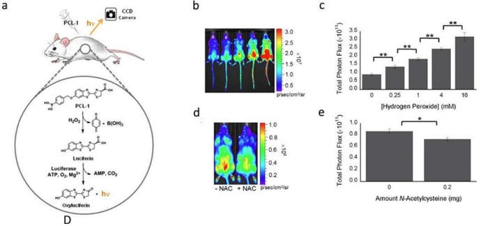
Source: Bioluminescence Imaging: Progress and Applications (NIH; 2011)
Medical Research
Bioluminescence has improved medical research, particularly using the imaging and detection techniques mentioned above.
One notable application is in cancer research, where bioluminescent markers help visualize and track tumor growth and metastasis in live animals. This allows researchers to monitor disease progression and evaluate the efficacy of treatments in real time. Bioluminescence is also used to detect tuberculosis (TB) and monitor oxidative stress in infertile men, providing noninvasive and highly sensitive diagnostic tools.
Drug Discovery and Development
Bioluminescence assays are widely used in drug discovery and development. They provide a rapid and sensitive means of screening potential drug candidates by measuring their effects on bioluminescent reporter genes.
These assays can identify compounds that modulate specific biological pathways, accelerating the identification of new therapeutic agents. Further, bioluminescence can be employed to monitor the distribution and efficacy of drugs within living organisms, enhancing the development of targeted therapies.
“Bioluminescence can be employed to monitor the distribution and efficacy of drugs within living organisms, enhancing the development of targeted therapies.”
Gene Therapy and Molecular Biology
Bioluminescence is also employed in gene therapy and molecular biology. Researchers use bioluminescent markers to track the expression and regulation of genes within live cells and tissues.
This application is crucial for studying genetic diseases and developing gene-based therapies. For instance, bioluminescent imaging helps scientists observe how genes are turned on or off in response to different treatments, providing insights into gene function and regulation.
Infection and Immunology
In the field of infection and immunology, bioluminescence enables the real-time tracking of pathogen spread and immune response. This is particularly useful in studying infections caused by bacteria and viruses.
Bioluminescent bacteria can be used to monitor infection progression and the efficacy of antimicrobial treatments. Similarly, bioluminescent markers help visualize the activity of immune cells, aiding in the understanding of immune responses to infections and autoimmune diseases.
Bioluminescence imaging faces several technical challenges, including limited tissue penetration and sensitivity to environmental conditions. False positives and negatives can occur due to inadequate signal detection or interference from other biological processes.
Standardizing and validating bioluminescence imaging techniques for clinical settings is essential to overcome these limitations and ensure accurate and reliable results.
The future of bioluminescence in medicine holds exciting possibilities. Researchers are exploring new ways to enhance the sensitivity and specificity of bioluminescent imaging techniques.
Advances in genetic engineering may lead to the development of new bioluminescent markers with improved properties, such as brighter and more stable luminescence. Additionally, integrating bioluminescence with other imaging modalities, such as MRI and PET, could provide more comprehensive diagnostic tools.
Another promising area is the application of bioluminescence in personalized medicine. By using bioluminescent markers tailored to individual patients, doctors could monitor the progression of diseases and effectiveness of treatments in real time, allowing for more precise and personalized therapeutic strategies.
This novel approach could enhance how diseases like cancer are treated, leading to better outcomes and improved patient care.
Bioluminescence represents a promising frontier in medical research and diagnostics. Its applications in imaging, drug discovery, and disease monitoring offer improved insights into biological processes and disease mechanisms.
While significant advancements have been made, there is still much to discover about the basic reactions and the full spectrum of bioluminescent systems. As research continues to evolve, bioluminescence is poised to play an increasing role in healthcare, paving the way for new diagnostic tools and therapeutic strategies.
"Bioluminescence represents a promising frontier in medical research and diagnostics. Its applications in imaging, drug discovery, and disease monitoring offer improved insights into biological processes and disease mechanisms."