


In the operating room, visiualization is critical. The success of a surgical procedure often hinges on the clarity with which the surgical team can see (and interpret) anatomical structures. Modern surgical lighting systems, like overhead lights, incavity lighting, and surgeon headlamps, work together to deliver optimized visual environments that optimize patient outcomes.
Effective lighting is essential for surgical safety, speed, and accuracy. Here, we examine how lighting impacts not only human vision but also machine vision in robotic and AI-assisted surgeries, highlighting emerging technologies that redefine what it means to “see clearly” in the operating room.
In any surgical setting - whether open, minimally invasive, or robotic - medical lighting must meet several demands at once:
Provide consistent, shadow-free illumination of the surgical site
Render tissue colors accurately to support real-time diagnostic decisions
Preserve depth perception for more precise and reliable spatial awareness
Avoid excessive heat or glare that can cause fatigue or interfere with procedures
These goals become challenging when lighting systems are obstructed by personnel, limited by anatomical constraints (e.g., deep cavities), or misaligned with the surgeon’s direct line of sight. In fact, 64% of surgeons report having to interrupt procedures to reposition lighting—an inefficiency that can contribute to delays, undue stress, and even critical errors.
Lighting also plays a key role in depth perception. Contour shadows - soft, angled shadows created by multi-source lighting - help surgeons distinguish depth and texture. Lighting systems that overcorrect for shadows may reduce depth cues, creating a flat visual field.
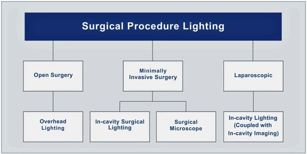
Source: Surgical Lighting: The Definitive Guide
An ideal system introduces just enough gradient to preserve spatial awareness while avoiding contrast shadows that obscure visibility. Well-designed surgical lighting offers:
Color fidelity, ensuring that tissue appears natural, aiding in the recognition of blood perfusion, fat layers, and pathologic margins, among other features.
Contrast and clarity to reduce the risk of misidentifying target tissues, imprecise dissection, and other forms of human error.
Even illumination, reducing eye strain, enhancing focus, and supporting surgeon endurance during complex and/or prolonged operations.
Here are some examples of well-known, commonly used medical lighting solutions that can be found in operating rooms worldwide.
Ceiling-mounted or boom-mounted surgical lights remain the primary source of general illumination in most operating rooms. Modern LED-based systems can deliver up to 160,000 lux with high color rendering indices (CRI 90+), including strong R9 values to accurately depict red tissue tones.
These systems illuminate the full surgical field and are designed to minimize contrast shadows - the harsh, obstructive shadows that occur when heads or hands block the light.
Despite these innovations, overhead lighting may struggle to reach into deep cavities or obscure corners, prompting the need for complementary solutions.
In modern surgical practices, in-cavity lighting plays a crucial role in enhancing precision and visibility during procedures. Utilized in both minimally invasive and open surgeries, this advanced technology delivers precise illumination directly into deep or narrow spaces through endoscopes, fiber-optic cables, illuminated retractors, or disposable LED strips.
In minimally invasive procedures, coaxial illumination aligns with the viewing axis to enable distribution of light without external obstructions. This can result in clear, shadowless images that aid surgeons in maintaining optimal visibility of surgical targets. In minimally-open procedures, in-cavity retractor-mounted lighting can diffuse shadows and bring the light where it’s most needed, clarifying anatomical features.
By minimizing deeper shadows and enhancing overall visibility, in-cavity lighting solutions significantly improve surgical outcomes, allowing surgeons to work with heightened precision and confidence.
“By minimizing deeper shadows and enhancing overall visibility, incavity lighting solutions significantly improve surgical outcomes.”
Surgeon-worn headlights are intensely focused light beams that follow a surgeon’s gaze. These are ideal for fine or deep work where external lights fall short. By being aligned with a surgeon’s direct line of sight, headlamps reduce shadows from hands and instruments.
However, ergonomic challenges persist, with 68% of frequent OR headlamp users reporting neck and/or back strain. Additionally, the field of illumination benefits only the wearer, while support staff still require additional lighting.
As robotic-assisted and computer-augmented surgeries become more common, medical lighting is no longer just for humans. Machine vision systems - whether embedded in surgical robots, endoscopic cameras, or AI-powered analysis tools - rely heavily on consistent, high-quality white light to function effectively.
Unlike human eyes, cameras and imaging algorithms are even more sensitive to inconsistencies in illumination.
They require consistent, high-CRI white light to:
Maintain sharpness and color accuracy in stereo or 3D endoscopic video feeds.
Provide clean inputs for tissue recognition, perfusion monitoring, or object tracking.
Reduce post-processing demands, such as white balancing or exposure correction.
Lighting systems with poor spectral balance or inconsistent intensity can degrade video quality and reduce the reliability of machine vision output. Let’s explore how lighting affects the performance of various machine vision systems in operating rooms.
In robotic-assisted procedures, stereo endoscopic cameras provide surgeons with a high-definition, 3D view of the surgical field. Advanced robotic platforms treat lighting as an integral part of the imaging chain, alongside camera sensors, image processing software, and display systems. In practice, bright, high-CRI white light enables these cameras to deliver accurate anatomical detail, support precise instrument tracking, and minimize the need for post-processing. Some robotic systems even prompt the surgeon to adjust lighting when image quality drops, while newer lighting systems can make adjustments automatically.
Many AI-driven tools now assist surgeons by analyzing surgical videos in real-time. These systems can track bleeding, assess perfusion, classify tissue types, or highlight anatomical landmarks. However, their performance is only as good as the images they receive, which depends entirely on lighting.
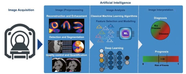
Source: European Journal of Nuclear Medicine and Molecular Imaging
High-fidelity lighting helps AI systems:
Detect subtle visual features, like bile staining, oxygenation levels, or tumor margins that require consistent color rendering.
Maintain calibration, which reduces the need for constant white balance or exposure correction in the video stream.
Interpret spatial relationships, especially in 3D reconstruction or motion tracking applications.
Newer LED surgical lighting systems with high R9 values (>70) ensure that red hues - critical for identifying blood, inflammation, and vascularity - are rendered vividly and consistently. This not only benefits human perception but also feeds better data into AI models.
Computer vision tasks, such as real-time 3D reconstruction, object segmentation, and motion tracking, are becoming 6 integral to image-guided surgery and autonomous robotics. These tasks require precise visual info, which in turn depends on structured, well-calibrated illumination.
Lighting considerations for computer vision include:
Controlled gradients: Slight variations in lighting across scenes can help algorithms extract topography and texture, akin to how human vision relies on contour shadows.
Avoiding glare: Overexposed regions or light reflected off metallic instruments can confuse segmentation algorithms or create false edges.
Shadow consistency: Too many harsh shadows can obscure features, but some shadows (like contour shadows) are useful for reconstructing depth.
For example, in procedures that involve tracking tool movements, high-frequency flicker-free lighting ensures video frames remain stable and usable for high-speed tracking. Some experimental lighting systems even dynamically tailor beam shape and intensity to match the movement of robotic arms or endoscopes, ensuring the area of interest is always optimally lit.
Proper lighting also plays a key role in training and deploying machine learning (ML) models.
Surgical AI systems are typically trained on datasets captured under specific lighting conditions. But if OR lighting deviates from those conditions, model accuracy can suffer.
Standardizing lighting conditions across different operating rooms is a critical part of model deployment. High-quality white light sources with a consistent spectrum and intensity enable AI models to operate across various environments with minimal recalibration.
Additionally, newer OR lighting systems integrate directly with endoscopic or overhead cameras, adjusting focus and beam shape based on where cameras are pointing, ensuring both human and machine vision are optimized simultaneously.
Whether through advanced overhead lights, targeted in-cavity systems, or AIpowered diagnostics, effective lighting is becoming part of a growing surgical ecosystem - one that interfaces with robots, cameras, and surgeon preferences in real-time.
In 2025 and beyond, surgical lighting will no longer be viewed as a passive utility. Intelligent LEDs, adaptive light shaping, wireless control, and integrated imaging will continue to transform the delivery and perception of light in operating rooms worldwide.
By investing in smarter, high-fidelity lighting systems, healthcare providers can support safer, faster, and more effective surgeries, while equipping clinicians with the visual and lighting solutions they need to deliver the best possible care.
“Newer LED surgical lightingsystems ensure that red hues arerendered vividly and consistently.”


Photodynamic therapy (PDT) is a light-activated, non-invasive treatment approach for certain cancers and skin conditions. Using a targeted combination of photosensitizing drugs and specific wavelengths of light, PDT triggers chemical reactions that selectively destroy harmful cells. In other words, it uses light not to see, but to heal.
Unlike traditional cancer therapies such as chemotherapy and radiation, which can carry heavy systemic side effects, PDT offers a highly localized, healthy tissuesparing solution. It is gaining traction in dermatology, oncology, and ophthalmology.
The precision of PDT makes it particularly compelling for therapeutic applications. By requiring both a drug and a specific light wavelength to activate treatment, clinicians can treat delicate or cosmetically sensitive areas without resorting to more invasive interventions.
At a very high level, PDT is a three-step process:
A patient is administered a photosensitizing agent, like a drug that is only absorbed by cancerous or abnormal cells.
The targeted area is exposed to a specific wavelength of light (usually in the redto-deep-red range of 600–800 nm).
This light activates the photosensitizer, generating reactive oxygen species (ROS) that damage or kill the target cells.
“PDT offers a highly localized, healthy tissue-sparing solution. It is gaining traction in dermatology, oncology, and ophthalmology.”
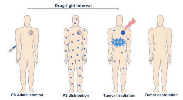
Source: Photodynamic Therapy Review: Principles, Photosensitizers, Applications, and Future Directions
Naturally, PDT is particularly effective for cancers or lesions located in or near the skin, or in organ linings that can be reached with light.
Beyond oncology, PDT is also being used to treat pre-cancerous conditions such as actinic keratoses, as well as non-malignant diseases like acne, psoriasis, Bowen’s disease, and lichen sclerosis.
Ongoing research is also evaluating the use of photodynamic therapy in metastatic or more challenging-to-reach cancers, utilizing more advanced light delivery systems.
Photodynamic therapy offers meaningful advantages for both patients and clinicians - advantages that extend well beyond tumor shrinkage or lesion clearance. By combining precision targeting with minimal invasiveness, PDT can deliver effective treatment outcomes with fewer risks, fewer complications, and better overall experiences.
Tissue-Sparing
Because photosensitizers accumulate primarily in abnormal tissue—and only become active when exposed to specific light wavelengths—PDT provides a level of spatial control that is difficult to achieve with chemotherapy or radiation. This makes it particularly valuable in cases where functional or cosmetic preservation is a priority, such as treating skin cancers on the face or lesions in the oral or ocular lining
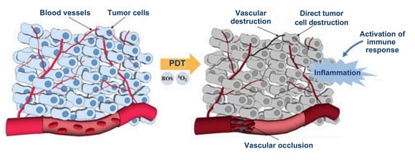
Source: Photodynamic Therapy Review: Principles, Photosensitizers, Applications, and Future Directions
Well-Tolerated
PDT is commonly well-tolerated, offering low systemic toxicity compared to traditional treatments. Patients avoid many of the harsh side effects associated with systemic drug exposure or radiation-induced damage, such as nausea, fatigue, immune suppression, or permanent tissue damage. The therapy is localized, and effects are mostly confined to the illuminated area.
Easily Repeatable
Unlike radiation, which can only be administered a limited number of times to the same area, photodynamic therapy can often be repeated at the same site if necessary, providing clinicians with more flexibility in treatment planning. The cumulative effect of these advantages often is a marked improvement in patient quality of life. PDT can lead to shorter recovery times, improved cosmetic outcomes, and more dignified care for individuals whose conditions might otherwise require invasive surgery or high-toxicity treatments.
Wavelength and Spectrum
PDT depends on precise optical parameters. Most photosensitizers have an absorption peak between 600 and 800 nm, corresponding to red or near-infrared light. Light above 800 nm does not carry enough energy to effectively excite oxygen and generate ROS, while shorter wavelengths do not penetrate deeply enough into tissue. ROS, Reactive Oxygen Species, are highly reactive molecules that help destroy targeted cells and signal the immune system to respond, making them essential to the therapeutic effect of PDT.
Light Intensity and Exposure
Achieving the right balance of fluence (energy per unit area) is critical. Too little light may lead to insufficient activation of the drug, while too much may damage healthy tissue or deplete tissue oxygen. Studies have shown that low fluence rates can be more effective because they prevent rapid oxygen depletion and support more complete photoactivation of the drug.
Color Temperature
While not often discussed in consumer terms, the color temperature of the light source must align with the absorption profile of the chosen photosensitizer. In practice, this means carefully tuning light-emitting devices - often via LEDs or lasers - to a narrow band of wavelengths for maximum therapeutic effect.
Heat and Risk Factors
Because light is being applied directly to human tissue, heat buildup must be managed. PDT devices are designed to emit “cold” light, minimizing tissue heating. Wearable or patch-based light delivery systems are under development to make treatment more comfortable.
Several promising technological innovations could expand photodynamic therapy across a growing number of medical applications beyond skin and surface-level tissues:
Improved photosensitizers: New generations of photosensitizing agents are being engineered with better photochemical properties, including stronger absorption in the therapeutic window (600–800 nm), increased selectivity for diseased tissues, and faster clearance from healthy tissue to reduce post-treatment photosensitivity.
Metal-Based Nanoparticles (MBNPs): Materials such as gold, silver, and titanium dioxide offer unique plasmonic and photocatalytic properties that enhance the activation of photosensitizers. By scattering and concentrating light at specific wavelengths, these nanoparticles can significantly enhance ROS generation. Early studies suggest that MBNPs could also enhance tumor selectivity and minimize off-target effects.
Wearable light-delivery devices: Innovations in wearable technology are also making PDT more accessible. Lightweight, battery-powered, or adhesive patchstyle devices are being developed to deliver consistent, low-intensity light for outpatient or even at-home therapy, especially for chronic dermatologic conditions or pre-cancerous lesions. These systems also increase patient comfort and compliance.
Enhanced treatment depth: Researchers are developing novel light delivery systems, including fiber optic probes, catheter-mounted LEDs, and interstitial illumination techniques. Additionally, formulation improvements in photosensitizers—such as those that target tumor vasculature or accumulate in hypoxic tumor cores—are extending the reach of PDT to more deeply embedded lesions.
Whole-body applications: Looking further ahead, there is growing interest in systemically administered photosensitizers combined with body-wide or organspecific light exposure. While still highly experimental, these approaches raise the possibility of PDT being used for metastatic disease or as a targeted adjunct to systemic therapies.
Photodynamic therapy exemplifies the power of light as a therapeutic tool—not just for visualizing disease, but for effectively treating it. As new technologies push the boundaries of what light can do—through advanced materials, smarter delivery systems, and deeper penetration—PDT is poised to play a larger role in cancer treatment and beyond.
It’s a vivid reminder that, in medicine, illumination and intervention are often one and the same.
“Several promising technological innovations could expand photodynamic therapy across a growing number of medical applications beyond skin and surface-level tissues”


Biophotonics is a rapidly advancing field that utilizes specific wavelengths of light to image, analyze, and manipulate biological tissue, resulting in more precise and less invasive diagnostics and therapies. It blends optics and nanotechnology to visualize structures in living systems, deliver drugs with pinpoint accuracy, and assist in tissue regeneration.
In 2025 and beyond, biophotonics is emerging as a very promising frontier in medical lighting and imaging. From detecting cancer with high-resolution optical imaging to guiding smart nanorobots deep within the body, it may offer very powerful tools to see and intervene.
Biophotonics represents more than a new toolset—it’s a new way of thinking about diagnostics and treatment. It enables clinicians and researchers to interact with the human body at the cellular and molecular levels, in real-time, with unprecedented precision.
One of the field’s most exciting contributions is the ability to visualize tissues with high resolution without the need for cutting, biopsy, or surgical intervention. Optical coherence tomography (OCT), photoacoustic imaging, and other modalities enable the visualization of tissue beneath the surface, often in vivo, facilitating faster and more accurate diagnoses.
“In 2025 and beyond, biophotonics is emerging as a very promisiging frontier in medical lighting and imaging.”
In therapeutic contexts, biophotonics enables light-guided targeting of diseased cells using nanoparticles or nanorobots, thereby minimizing damage to surrounding healthy tissue. These agents can be activated with specific wavelengths of light, releasing drugs or triggering cell destruction only where needed.
As with photodynamic therapy (PDT), the non-invasive nature of these interventions results in reduced systemic toxicity compared to traditional chemotherapy or radiation. This opens the door to more tolerable, repeatable treatments that patients can undergo with fewer side effects.
Biophotonic tools also may enable continuous or periodic monitoring of chronic conditions via embedded or wearable sensors. These platforms can provide realtime insights into biological markers, such as pH, oxygenation, and inflammation, enabling easier management of complex diseases without the need for repeated hospital visits.
Finally, biophotonics is creating new possibilities in tissue regeneration and repair. By stimulating biological processes with light or delivering regenerative agents via light-responsive nanocarriers, researchers are harnessing biophotonics actively to support healing at the microscopic level.
The biophotonics field depends on smart material design and precise optical engineering. Key technical components include:
Wavelengths and Penetration: Most imaging and treatment applications rely on near-infrared (NIR) and near-infrared-II (NIR-II) light (650–1700 nm), which penetrate tissue deeply while avoiding interference from autofluorescence and hemoglobin absorption.
Light Intensity and Exposure: Power density and exposure time must be carefully adjusted to optimize signal clarity and therapeutic efficacy while minimizing phototoxicity and tissue overheating.
Nanorobot Design: Nanorobots and nanoparticles are engineered for biocompatibility, surface stability, and clearance. Many platforms utilize biodegradable materials or rely on the rapid renal excretion of unbound agents.
Heat and Toxicity Risks: Photothermal therapies and quantum-dot–based agents require careful control to avoid overheating and minimize exposure to potentially harmful heavy metals. Efforts are underway to develop safer, metalfree alternatives.
At its core, biophotonics uses light-responsive materials, often in combination with nanoparticles or nanorobots, to interact with tissues in ways that traditional imaging or drug delivery methods cannot.
“Biophotonics is creating new possibilities in tissue regeneration and repair. Researchers are harnessing biophotonics actively to support healing at the microscopic level.”
Biophotonic Imaging
Techniques such as luminescence, photoacoustic imaging (PAI), surface-enhanced Raman scattering (SERS), and optical coherence tomography (OCT) allow for detailed, non-invasive visualization of biological structures and molecular signals.
By operating in NIR and NIR-II windows (650–1700 nm), these imaging systems achieve deeper tissue penetration while minimizing interference from endogenous fluorophores, such as hemoglobin. This is especially useful for early disease detection, intraoperative guidance, and real-time monitoring.
Targeted Light Therapy
Biophotonic systems can also be used to treat disease, not just detect it. Fluorescent nanoparticles and light-activated nanorobots can be engineered to respond to external wavelengths, triggering localized therapeutic responses only when and where needed.
These include PDT, which generates reactive oxygen species (ROS) to destroy abnormal cells; photothermal therapy (PTT), which uses focused heat to ablate tissue; and light-triggered drug release, which delivers therapeutics directly to affected sites. The result is more targeted treatment with fewer side effects.
Bio-Detection Diagnostics
Biophotonic probes—including red blood cell–based microlenses, bioluminescent agents, and biologically inspired waveguides—enable the sensitive detection of biological changes, such as blood pH, inflammation, or the presence of infectious agents.
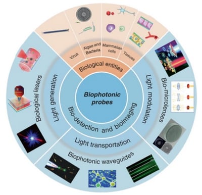
Source: Biophotonic probes for bio-detection and imaging
These probes can provide clinicians with real-time diagnostic data at the point of care, aiding faster decision-making for conditions ranging from cancer to sepsis to cardiovascular disease. Many of these systems are designed for integration with wearable or implantable devices, leading to continuous, personalized monitoring.
The impact of biophotonics spans across a wide spectrum of medical disciplines, offering new tools for diagnosis, targeted therapy, and real-time monitoring in ways that are reshaping clinical possibilities:
Oncology: Biophotonics is playing a pivotal role in cancer care, from highresolution imaging of tumor margins to light-triggered delivery of chemotherapeutics. Nanorobots activated by light or magnetic fields are being explored for targeted ablation of tumor cells—offering precision therapy with reduced systemic toxicity.
Neurology: In the growing field of neurophotonics, researchers are developing ways to “probe the brain without probing the brain.” Applications include the use of light to study and treat chronic pain, Alzheimer’s, Parkinson’s, and psychiatric disorders through controlled neural modulation and optical biomarkers.
Cardiology: Light-powered micro- and nanorobots are being developed for intravascular use, including precise delivery of thrombolytic agents to dissolve clots and targeted therapy for atherosclerotic lesions. Biophotonic sensors can also track vascular inflammation, plaque development, and blood chemistry in real-time.
Infectious Diseases: In vivo biophotonic imaging allows researchers to track the progression and resolution of infections in animal models without invasive sampling. This has already led to improved understanding of pathogen behavior, host immune response, and vaccine efficacy.
Ophthalmology: Light-activated drug delivery systems and diagnostics are opening new frontiers in eye care, particularly for diseases like macular degeneration, diabetic retinopathy, and uveitis. Biophotonic systems are being engineered to target intraocular tissues selectively, enabling localized treatments.
Personalized Medicine: By tailoring therapies to a patient’s unique optical and biological profile - for example, tracking tumor phenotypes, monitoring immune activity, or dynamically adjusting drug dosing—biophotonic systems enable a new level of individualized, responsive care across medical fields.
“The impact of biophotonics spans across a wide spectrum of medical disciplines, offering new tools for diagnosis, targeted therapy, and real-time monitoring in ways that are reshaping clinical possibilities.”
As biophotonics continues to evolve, its role is expanding from isolated diagnostic or therapeutic functions to more integrated, intelligent systems that can navigate complex biological environments with minimal disruption.
Optical Waveguides
To overcome the depth limitations of surface-applied light, researchers are developing new classes of implantable or biodegradable optical waveguides. These structures can channel light deep into tissue with low loss, enabling targeted therapies in organs and systems that were previously inaccessible by non-invasive methods.
Materials such as silk fibroin, calcium-phosphate glass, and genetically engineered proteins are being engineered for both optical performance and biological compatibility.
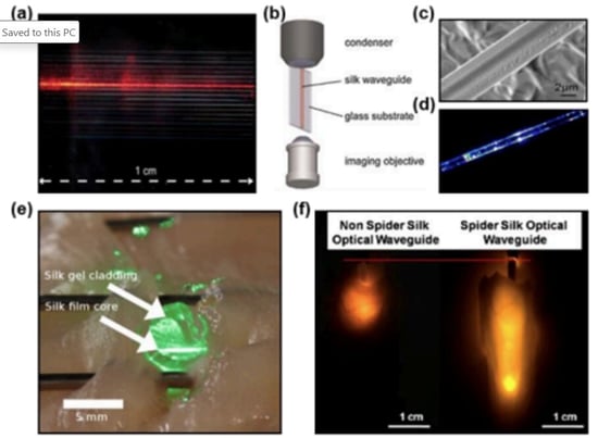
Figure 3. Waveguides made of natural materials. (a) Silk optical waveguide. Reproduced with permission [34]. Copyright 2010, Wiley-VCH. (b) Tools to analyze transverse faces of waveguides. Reproduced with permission [34]. Copyright 2010, Wiley-VCH. (c) Scanning electron microscopic (SEM) image of a silk fiber. Reproduced with permission. [54] Copyright 2013, AIP. (d) Micro-beam profile of a spider silk fiber. Reproduced with permission. [54] Copyright 2013, AIP. (e) An implanted silk optical fiber in tissue. Reproduced with permission. [51] Copyright 2015, OSA. (f) Comparison of light penetration in diferent waveguides. Reproduced with permission [55]. Copyright 2017, ACS.
Source: Biocompatible and Implantable Optical Fibers and Waveguides for Biomedicine
Waveguides can also be paired with sensors or actuators, opening up new avenues for smart implants that deliver light and feedback in real-time. .
Biophotonic Nanorobots
Nanorobots represent a cutting edge of light-activated therapy and diagnostics. These autonomous or remotely guided micro-machines are being designed to navigate biological terrain, respond to molecular cues, and deliver payloads with pinpoint accuracy.
In cancer care, they are being tested for mitochondrial targeting, enhancing the precision and potency of drug delivery while reducing collateral damage. In neurology, their ability to cross the blood-brain barrier could significantly advance how we treat brain tumors, neurodegenerative diseases, or even psychiatric conditions.
Future nanorobots may combine sensing, analysis, and actuation within a single platform—essentially becoming mobile “labs” inside the body.
AI/ML Integrations
The fusion of biophotonics with artificial intelligence and machine learning (AI/ML) is enabling the development of more intelligent and adaptive systems. Algorithms are being used to interpret biophotonic imaging data, guide nanorobot navigation, optimize therapeutic dosing, and even predict patient responses in real-time.
For instance, AI-driven pattern recognition can enhance image segmentation, detect early signs of pathology, or help nanorobots avoid healthy tissue. These systems are paving the way for semi-autonomous interventions that combine clinical oversight with algorithmic precision, especially useful in complex or rapidly evolving clinical situations.
Neurophotonics and Pain Management
In the brain and nervous system, biophotonics opens new frontiers in non-invasive neural modulation. Light can be used to alter neural circuit activity without penetrating the skull, offering potential alternatives to electrical implants or pharmacologic intervention. Clinical targets include chronic pain, epilepsy, depression, and neuroimmune disorders.
By targeting light-sensitive opsins, proteins found in the retina of the eye, or leveraging thermal and photoacoustic effects, neurophotonics may eventually allow for precise, personalized control of neural function, guided by closed-loop feedback from biophotonic sensors.
Biophotonics represents a big step forward in the evolution of medical lighting, from passive visualization to active intervention. By combining high-resolution imaging with light-triggered therapy and smart nanoscale engineering, this exciting field can unlock new possibilities for early detection, precise treatment, and real-time monitoring.
Whether enabling a blood test that maps disease with light or guiding a nanorobot through the brain, biophotonics exemplifies what is possible when light becomes not just a source of illumination, but a tool for effective and sustainable healing.
Biophotonics represents a big step forward in the evolution of medical lighting, from passive visualization to active intervention.


Dialysis has remained largely unchanged for over six decades. Despite incremental advances in machine design and home hemodialysis, most patients with end-stage renal disease (ESRD) still rely on large, immobile machines and time-consuming treatment schedules—often three times per week, several hours at a time.
An emerging technology called photooxidation-based dialysis could upend this model. By using targeted light to break down urea, the most abundant nitrogenous waste product in the blood, photooxidation could enable the continuous recycling of dialysate fluid, significantly reducing the size and complexity of dialysis machines.
In doing so, photooxidation-based dialysis could also pave the way for wearable, portable, or home-based dialysis solutions that better align with patient lifestyles.
The core innovation behind photooxidation-based dialysis is how it decomposes urea using light-driven chemical reactions.
Unlike conventional systems that require large volumes of water to flush toxins from spent dialysate, photooxidation technology can break down urea into harmless nitrogen and carbon dioxide gases vented from the device. The cleaned fluid can then be cycled back into the machine for reuse.
“Photooxidation-based dialysis could also pave the way for wearable, portable, or home-based dialysis solutions that better align with patient lifestyles.”
This closed-loop regeneration process significantly reduces the fluid volume required for each treatment, enabling smaller and more mobile machines. A wearable system using this technique could theoretically run continuously or semi-continuously, more closely mimicking the natural function of healthy kidneys.
The ultimate goal is a dialysis device small enough to be worn like a backpack, freeing patients from the physical and logistical burdens of in-center treatments. Photooxidation may one day allow dialysis to shift from a disruptive, scheduledefining burden to a background therapy that patients can carry with them.
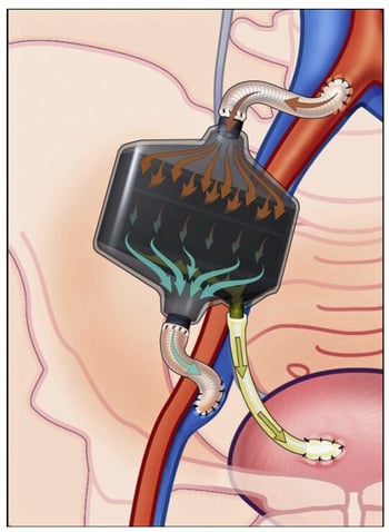
Figure shows a portable, wearable and implantable artificial kidney systems; however, their implementation has manufacturing, feasibility and logistics challenges.
Source: From Home to Wearable Hemodialysis: Barriers, Progress, and Opportunities
More than 500,000 people in the U.S. currently rely on dialysis. For many, access to portable or wearable systems would represent a great improvement in how they live, work, and manage their health.
The implications of this groundbreaking technology are significant:
Enables mobility: By eliminating the need for external water systems and large dialysate reservoirs, photooxidation opens the door to ambulatory dialysis treatments. Patients could receive therapy while going about their daily lives—at work, on errands, or even while traveling—without being tethered to a clinic or home station.
Improves quality of life: Patients would no longer need to schedule their lives around in-center dialysis appointments multiple times a week. Instead, they could treat themselves on their own time, reducing the disruption to personal and professional commitments.
Reduces infrastructure burden: Smaller, wearable dialysis systems could significantly ease the load on healthcare infrastructure. Clinics and hospitals would see decreased demand for dialysis chairs, machines, and staff time— valuable in high-demand urban settings or resource-constrained areas.
Incorporates patient-first design: Research teams are prioritizing humancentered design. Key considerations include lightweight, backpack-sized devices for enhanced wearability, proprietary connection systems that eliminate the need for large-bore needle sticks, and modular, disposable cartridges that simplify setup, maintenance, and replacement.
Roadblocks to Portable Dialysis
Key engineering and biomedical challenges remain. One key hurdle is designing a dual-loop system that keeps the high-pH environment necessary for photooxidation separate from the patient’s blood-contacting components. This architectural separation is crical to prevent biocompatibility issues while ensuring efficient toxin removal.
Material safety also poses a significant challenge. Some early photoelectrode designs are prone to silver ion leaching, which can be toxic in the long term. Likewise, while photooxidation has been shown to effectively decompose urea into benign gases, some studies report the presence of small but persistent levels of nitrite accumulation.
Finally, portability demands not only miniaturization but also safe, reliable operation in changing real-world environments. Engineers must design lightweight, durable, energy-efficient, and easy-to-operate systems, without compromising treatment performance or safety.
“Patients would no longer need to schedule their lives around incenter dialysis appointments multiple times a week. Instead, they could treat themselves on their own time.”
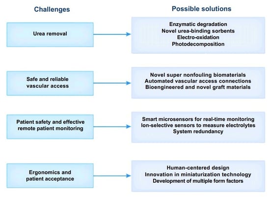
Figure 2. Key challenges and possible solutions for successful development of wearable dialysis technologies.
Source: From Home to Wearable Hemodialysis: Barriers, Progress, and Opportunities
Photooxidation-based dialysis is still in its early stages, but is already gaining attention. By offering a non-mechanical, chemical-free method to break down urea in real time, the technology stands apart from conventional filtration- or sorbent-based dialysis systems.
One advanced prototype is the AKTIV (Ambulatory Kidney to Increase Vitality), developed by the Center for Dialysis Innovation at the University of Washington. This portable system utilizes ultraviolet light to decompose urea into nitrogen and carbon dioxide, allowing for the continuous regeneration of dialysate without the need for an external water supply.
Although still experimental, AKTIV has demonstrated proof of concept with 24-hour operation in laboratory conditions. Importantly, it achieves this using low-power, commercially available LED sources in the 365–460 nm range, suggesting strong potential for scalability and cost-effectiveness.
The AKTIV concept integrates multiple modules, including a blood management cartridge, a dialysate regeneration chamber, and a smart control unit—designed to fit into a lightweight, wearable format. The goal is to create a safe, portable, closedloop dialysis system.
Recent studies have also validated the potential of photooxidation-based urea decomposition:
Wavelength and Intensity: Effective decomposition has been achieved using light wavelengths between 365 and 460 nm and power densities around 18 mW/cm², making the process compatible with modern LED technology.
Urea Removal: Photocatalytic materials, such as titanium dioxide (TiO₂) and hematite (Fe₂O₃), especially when coated with nickel oxyhydroxide (NiOOH), have demonstrated the ability to break down therapeutic quantities of urea over a 24-hour period. In one model, just 0.5 m² of photocatalytic surface was sufficient to clear a full day’s urea production in a typical patient.
Selectivity and Safety: Photoxidation can offer a safer profile compared to electrooxidation, which can generate harmful by-products like chloramines and chlorine gas. Studies show minimal or no production of toxic byproducts, such as ammonia or nitrate, although some formation of nitrite has been observed and requires further refinement.
Bringing photooxidation-based dialysis from prototype to clinical use will require cross-disciplinary breakthroughs in material science, photochemistry, biomedical engineering, and patient-centered design.
While the underlying science is sound, translating lab success into everyday medical practice demands real-world optimization across multiple fronts:
Refining photoreactor efficiency: Next-generation devices must maximize urea breakdown within a compact footprint by increasing the effective surface area of photocatalytic materials, improving light absorption and photon utilization, and ensuring long-term catalyst stability under repeated use. Researchers are exploring advanced geometries, nanostructured coatings, and improved LED integrations.
Dual-loop system architecture: Because photooxidation requires a high-pH environment for optimal chemical reactions, it cannot directly interact with blood-contacting components. One promising solution is a ‘dual-loop’ system that has a patient-safe dialysate loop for blood exchange and a separate, sealed photooxidation loop for urea removal.
Human-centered design: Mobility is only part of the equation. Patients and caregivers also require simplicity, comfort, and confidence in use. The physical and emotional experience of wearing the device should be frictionless. Incorporating feedback from real patients will be crucial to long-term adoption.
Photooxidation-based dialysis represents a significant and promising advancement in renal care. By using light to regenerate dialysate in real time, this technology could dramatically reduce the size of dialysis machines—freeing patients from clinical chairs and giving them back control over their time, lifestyle, and mobility.
Though it’s still in its early days, the convergence of photochemistry, smart materials, and patient-driven design suggests a future of wearable, home-based dialysis may be closer than we think. At the heart of that future is a deceptively simple idea: that light can carry the burden that water once did—cleansing, renewing, and enabling life on the move.
“Recent studies have validated the potential of photooxidation based urea decomposition.”