


.svg)
Some of the hardest places to illuminate in surgery are also some of the smallest. A deep breast pocket. A narrow spinal corridor. A subcutaneous tunnel. A confined space beneath a tissue flap.
In these environments, the challenge is not simply producing more light. It is getting useful light to tissue at depth while occupying as little of the surgical working space as possible.
That is changing how surgical lighting is designed. Instead of treating illumination as something separate from the instruments creating access to the surgical field, light is moving closer to the anatomy itself. Retractors, fiber-optic light guides, flexible optical components, and increasingly integrated surgical instruments can deliver illumination from within the working space.
At the same time, optical components are becoming smaller and more capable. Together, those trends are pushing in-cavity lighting toward a different design goal: not maximum brightness, but precise illumination delivered exactly where it is needed.
Deep surgical fields create an optical problem that becomes more difficult as the access point gets smaller.
Light has to reach tissue through an opening already occupied by retractors, instruments, the surgeon's hands, and surrounding anatomy. The deeper and narrower that space becomes, the more important the location and distribution of the light become.
Research using a simulated pelvic wound demonstrated the effect. Knulst and colleagues compared different approaches to illuminating a difficult-access surgical cavity. Locally positioned prototype light sources delivered 12.2 lumens inside the wound, compared with 5.7 lumens from a head-mounted source. The locally positioned sources also distributed light across a substantially wider range of angles inside the cavity (Knulst et al., 2011).
The takeaway is not that one particular device is the answer. It is that location fundamentally changes light delivery.
Moving the illumination closer to the tissue can allow more of the available light to reach the areas the surgeon is actually trying to see.
For in-cavity lighting, the important question is not "How much light does the source produce?" It is "How much useful light reaches the tissue?"
Moving Light Into the Surgical Field
In-cavity lighting applies that idea by bringing the point of illumination inside the working space.
A fiber-optic light guide can be incorporated into a retractor. A flexible optical element can run along an instrument. Light can be delivered near the distal end of a device rather than from a separate source positioned farther away.
Several surgical specialties demonstrate the same underlying design principle.
In breast surgery, lighted retractors have been used to provide illumination beneath tissue during procedures performed through limited access points. Zhu and colleagues documented their use during breast-conserving surgery through a peri-areolar incision (Zhu et al., 2019).
In orthopedic surgery, Broderick and colleagues described a mini-open fasciotomy performed through a 3 to 4 cm incision. A fiber-optic lighted retractor was advanced along the subcutaneous plane, providing direct visualization of the fascia and superficial peroneal nerve within the narrow working corridor (Broderick et al., 2020).
An illuminated retractor has also been evaluated in cochlear implantation, where visualization is required inside a confined subperiosteal pocket. In a retrospective study of 117 cases, its use did not produce a statistically significant reduction in total operative time, although no adverse events were attributed to the device (Alrashidi et al., 2017).
That is a useful reminder not to overstate the benefit. In-cavity illumination does not automatically make every procedure faster. Its more fundamental purpose is to make useful light available in anatomy that is difficult to illuminate.
Bringing illumination into the surgical field creates a second challenge: there is very little room for the lighting system itself.
Every component placed within a narrow access channel competes for working space. That puts pressure on lighting systems to become thinner, lower-profile, and more integrated with instruments already required for the procedure.
Miniaturization in this context does not simply mean making a smaller light source. It means reducing the physical footprint of the entire optical delivery system while maintaining adequate illumination, distribution, color quality, durability, and thermal performance.
Recent developments elsewhere in biomedical optics show how quickly the underlying technology is progressing.
In 2025, researchers at Carnegie Mellon University reported a flexible thin-film optical device measuring only 7 × 400 μm and incorporating multiple parylene photonic waveguides for light delivery and collection from deep tissue (Malekoshoaraie et al., 2025). The device is an experimental imaging system, not a surgical retractor, but it illustrates how thin and flexible optical delivery structures are becoming.
In 2026, another optical design study explored miniaturized multimodal endoscopic systems using scanning fibers and wavelength-selective diffractive optics to handle multiple spectral ranges in a compact architecture (Stumpf et al., 2026).
Neither technology is a replacement for today's in-cavity surgical lights. What they demonstrate is a broader trend in medical photonics: sophisticated optical functions no longer require large optical assemblies.
For surgical device designers, that creates new possibilities for putting illumination into places where there was previously little room for it.
Miniaturization creates opportunities, but it also makes the engineering problem harder.
Distribution Matters
A small, bright point source can produce hotspots without adequately illuminating the rest of the field. Light has to exit the device in a pattern appropriate for the anatomy.
Depending on the procedure, that could mean forward illumination, a broad lateral distribution, or light emitted along a portion of the instrument.
This is why the Knulst study is particularly relevant. The locally positioned prototypes improved not only the amount of light entering the simulated wound, but also the angular distribution of that light (Knulst et al., 2011).
The goal is not simply more light in less space. It is controlled light in less space.
Color Still Matters
Brightness alone does not determine what a surgeon can see. The spectrum of the illumination affects how tissue appears and how easily subtle color differences can be recognized.
Mundinger and Houser asked 16 surgeons across 11 specialties to evaluate vasculature, colon, omentum, liver, and bone under approximately 3000 K, 4000 K, 4500 K, and 5100 K illumination. The 3000 K condition was rated lowest, while no significant differences were found among 4000 K, 4500 K, and 5100 K. The study also found no evidence that one spectrum was consistently better for a particular tissue or surgical specialty (Mundinger & Houser, 2019).
The practical takeaway is not that higher color temperature is always better or that every surgical light needs adjustable settings. Rather, a neutral-to-cool white spectrum in the 4000 K to 5100 K range may provide a more dependable starting point than warmer illumination. For color-dependent tasks, such as distinguishing vasculature from surrounding tissue, assessing perfusion, or recognizing tissue boundaries, the spectrum should be evaluated against the actual anatomy and procedure rather than selected by color temperature alone.
As optical systems become smaller, maintaining reliable color rendering and appropriate spectral performance remains part of the design challenge.
Heat Does Not Disappear With Size
Miniaturization can also make thermal performance more important because optical energy and heat are being managed within a smaller physical area.
A 2022 study of fiber-optic lighted retractors in breast surgery illustrates how much system design matters. Researchers compared 3.0 mm and 5.5 mm optical cables used with the same type of lighted retractor.
The 5.5 mm cable reached a peak connection temperature of 49.23°C. The 3.0 mm cable peaked at 33.29°C. Yet measured illumination was almost identical at approximately 2,450 and 2,500 lux, respectively (Nizzero et al., 2022).
In other words, a larger optical component did not deliver meaningfully more measured light in that setup, but it did produce considerably more heat at the connection.
Optical efficiency, coupling, materials, geometry, and thermal management all have to be considered as a system.
Lighting Is Becoming Part of the Instrument
Miniaturization becomes especially powerful when lighting no longer needs its own space at all.
A recent example comes from neurosurgery. Lavrador and colleagues reported a 2024 single-center study of an instrument incorporating an in-situ light source near the working tip. The system could deliver white or 405 nm blue light within approximately 15 mm of tissue while also functioning as a microdebrider.
.png?width=744&height=655&name=image%20(8).png) Figure 1. Instrument-integrated light delivery within a constrained surgical corridor. The panels show the microdebrider tip with an adjacent light source, light source positioned beside instrument’s lateral cutting edge and assembled system inside a tubular retractor
Figure 1. Instrument-integrated light delivery within a constrained surgical corridor. The panels show the microdebrider tip with an adjacent light source, light source positioned beside instrument’s lateral cutting edge and assembled system inside a tubular retractor
The study included 35 patients, eight of whom underwent tubular retractor-assisted minimally invasive surgery (Lavrador et al., 2024). The application is highly specialized, and the study represents early clinical experience rather than evidence that the technology should be generalized across surgery.
But the architecture is significant. Light delivery is becoming another function that can be engineered into the surgical instrument alongside retraction, cutting, suction, navigation, imaging, or sensing.
That changes the role of illumination. Instead of adding another device to an already crowded surgical field, the lighting system can begin to disappear into the instruments that are already there. The ultimate form of miniaturization may be lighting that takes up almost no additional surgical space because it has become part of the instrument itself.
There is unlikely to be one ideal in-cavity lighting design. A breast pocket, spinal corridor, orthopedic tunnel, and neurosurgical access channel have different depths, geometries, tissue types, and instrument requirements.
That means the next generation of in-cavity illumination may become increasingly procedure-specific. The questions become more precise:
These are optical and mechanical design questions as much as they are lighting questions.
.png?width=2400&height=1350&name=image%20(9).png) Figure 2. Similar light output does not guarantee similar thermal performance.
Figure 2. Similar light output does not guarantee similar thermal performance.
Several directions are beginning to converge:
Taken together, these developments point toward a surgical lighting model that is smaller, more targeted, and more closely integrated with the procedure itself.
Put Light Where the Work Is
The future of in-cavity surgical lighting is not simply brighter. It is closer, smaller, and more precise.
As surgical access becomes more constrained, lighting has to do more with less space. That shifts the engineering challenge away from raw output and toward delivered illumination: where the light originates, how it spreads, how accurately it renders tissue, how much heat it creates, and how seamlessly it fits into the instrument.
The most effective in-cavity lighting may ultimately be the lighting surgeons hardly notice as a separate system at all. It is simply part of the tool, delivering the right light to the right anatomy at the right time.
-2.png)
For most of medicine’s history, good clinical light has been hard to come by. A wall outlet, a ceiling mount, and a fiberoptic cable running back to an external box. Wherever the power was, that was where the light stayed. That is where care had to happen.
Battery-powered medical lighting reimagines medical lighting for the modern world. By moving the energy source onto the device itself, rechargeable lithium-ion or lithium-polymer cells powering high-efficiency LEDs, a whole category of lights now delivers clinical-grade illumination with no cord, no box, and no dependence on the room where it is used.
It may sound like a convenience, but portable medical lighting is so much more than that. Untethering light sources also untethers care: surgical-grade illumination can reach a field hospital, a disaster zone, a patient’s bedside, or their skin.
And because the same battery-and-LED platform that lights a surgical cavity can also deliver therapeutic light, portable systems are the rare device category that does double duty, helping clinicians see while helping patients heal.
Untethering light sources also untethers care: surgical-grade illumination can reach a field hospital, a disaster zone, a patient’s bedside, or their skin.
Fixed lighting infrastructure solves one problem well: brightness in a known location. Hang a luminaire array over an operating table, and you get reliable, high-output light exactly where the table sits. The trouble starts the moment care needs to move, or the moment the light needs to reach somewhere a ceiling-mounted beam cannot.
Those limits recur across settings. Cables tether the clinician to a box, creating trip hazards on the sterile floor and adding to setup time. Overhead luminaires light the field from above and behind the surgeon, so the deeper and narrower the surgical pocket, the more the surgeon’s own hands and instruments block the beam.
In more limited settings—including field hospitals, mobile units, rural clinics, and disaster zones—there is often no reliable power, so there is no fixed lighting. Sterility is a quieter liability: reusable corded components are reprocessed between uses, and every reusable part that crosses the sterile field is another potential point of failure to clean and track.
Instead of drawing power from a fixed main supply, portable lighting devices carry their own power sources. Rechargeable cells drive high-efficiency LEDs for stable brightness, adjustable beam geometry, and extended runtime in a compact, cablefree package. Many designs add status signaling (e.g., battery level, mode changes) so the clinician always knows the device state.
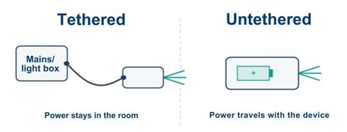
Figure 1. Tethered versus untethered lighting. In tethered lighting models, the light source draws power from a fixed power source. Battery-powered designs place the energy directly on the device, and the light travels with it.
Currently, the category is broad: surgical headlamps, handheld and stand-mounted exam lights, mobile surgical field lights, lighted retractors, and wearable therapeutic light sources. What unifies them is not a shared application but a shared principle, power autonomy:
Power and endurance: Hot-swappable battery modules keep a light running through long procedures without interruption. Charge monitoring is not optional. A portable light without a real-time state-of-charge display risks failing midprocedure, so intelligent control circuits with charge monitoring and external charging capability are essential.
Brightness and optics: High-power LED headlamps reach tens of thousands of lux at typical working distances, with commercial surgical headlights commonly delivering 15,000–30,000 lux, depending on the selected runtime mode. In practice, most portable lights serve as supplemental sources, filling shadows and reaching cavities that overhead arrays cannot, with adjustable spot size and focal length as standard features.
Color temperature and rendering: When surgeons evaluated LED spectra across the 4000–5100 K range, they rated all spectra as effective, with no significant differences, while a warmer 3000 K spectrum was rated lower. The IEC specifies an acceptable range for surgical lighting of 3000–6700 K, with a color-rendering index of at least 85. The same evaluation found statistically significant “spectrum-by-surgeon” preferences, meaning there is no single best color temperature for every operator.
Heat and risk factors: Thermal load is the constraint that scales worst, and more wattage does not automatically buy more useful light. Past a point, added power mainly adds heat, which is why thermal management matters as much as raw output. Engineering responses include heat sinks, liquid cooling, and moving the battery to a waist pack to shift thermal load off the head.
Sterilization and durability: Batteries and electronics cannot survive an autoclave, which forces a design choice. Detachable sterile coverings, sterilizable handles kept separate from the light engine, or fully single-use disposables?
Where fixed lighting is defined by location, portable lighting is defined by its reach. The same untethered platform appears in nearly every setting where care is delivered.
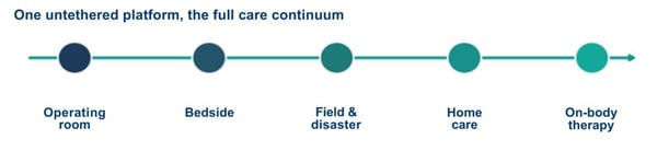
Figure 2. One untethered platform across the care continuum. The same battery-and-LED approach that supplements an operating room also serves the bedside, the field, the home, and on-body therapy.
The operating room: Battery-powered headlamps, handheld field lights, and cordless lighted retractors supplement or replace overhead illumination, bringing light into deep pockets without cables crossing the sterile field. Here, the defining trait is the power source, not the instrument design. The same retractor that another team integrates light into is simply one more device freed from the wall.
The bedside: Handheld and stand-mounted exam lights reveal surface detail, color change, and bleeding patterns that ambient room light washes out, supporting wound assessment and minor procedures for hospital inpatients.
Field, disaster, and military medicine: In austere settings with no fixed power, battery-powered systems are not a convenience but the only option. Military medical support in particular is a pressing need, and recent design work proposes modular, compact lighting that one person can carry to set up a workspace where none existed.
Home and continuous care: Untethered light follows patients out of the clinic, into post-operative monitoring, chronic wound care, and disease management performed in living rooms rather than exam rooms.
Therapeutic delivery: Battery-powered photobiomodulation (PBM) devices— including handheld units, wearable patches, and light-emitting bandages— deliver specific therapeutic wavelengths directly to tissue, a role no fixed luminaire can fill.
The current generation of portable lighting devices is a starting point, but several developments are converging on even smaller, smarter, and more portable light sources. As these devices mature, therapy that once required a clinic visit could run continuously at home.
Flexible, skin-conformable form factors are maturing fast: luminous fabrics that hold stable output over hours of wear with confirmed biocompatibility, OLED-based patches that deliver uniform, low-heat irradiation, and combinatorial dressings that pair nanofibrous wound material with flexible LED arrays. A wearable platform that combines vital-sign sensing with PBM has even been proposed for early treatment of traumatic brain injury in military settings.
The hardest lighting environments—including front-line, disaster, and resourcelimited settings—have historically been an afterthought in luminaire design. That is changing. Recent work directly addresses the lack of specialized lighting for these settings with modular, transferable systems that can be carried, assembled quickly, and run on batteries.
Gesture-recognition controls, the subject of recent patent activity, let a surgeon adjust a sterile headlamp without breaking scrub. (Paul et al., n.d., NIH.gov) Sensorand AI-driven systems aim further ahead, toward optimized surgical lighting that adapts its intensity to the procedure phase, ambient conditions, and the battery state on its own.
Push power autonomy to its smallest scale, and you arrive at fully implantable, wirelessly powered light sources, the domain of optogenetics, where micro-LEDs weighing a fraction of a gram deliver light inside neural tissue. It’s the same engineering problem as a cordless headlamp (miniaturized power, thermal control, biocompatible packaging).
For decades, a patient’s lighting quality depended on the quality of the room they were in. Fixed infrastructure meant fixed geography with fixed limitations. The best medical illumination lived in the best-equipped operating rooms. Everywhere and everyone else had to make do.
By carrying their own power, portable lights bring clinical-grade illumination to the bedside, the field, the disaster zone, and the home. They also increasingly deliver light-based therapies, not just improved visibility.
As batteries shrink, optics sharpen, and controls grow smarter, the question is no longer where good medical light can reach, but rather where and how we decide to deliver it.
For decades, a patient’s lighting quality depended on the quality of the room they were in. Fixed infrastructure meant fixed geography with fixed limitations. The best medical illumination lived in the best-equipped operating rooms.


With six decades of research behind it, Photobiomodulation (PBM) should be a breakthrough therapy. PBM has proven to be non-invasive, well-tolerated, with a compelling cellular mechanism: specific wavelengths of red and near-infrared light are absorbed by mitochondria, accelerating production of ATP, helping cells repair, reduce inflammation, and pain.
By every obvious measure, PBM should be widely practiced everywhere. But it is not. So the question the field keeps asking is fair: Why hasn’t PBM become standard-of-care in widespread applications?
The answer is complex, but the pathway has begun to clear. In November 2024, the FDA authorized the first device of its kind, and several bottlenecks that have held PBMs back for half a century are beginning to give way.
In this article, we will take a look at where PBM has earned its place, what has kept it from spreading further, and what is finally starting to change.
Before turning to why PBM has lagged, it helps to be precise about what it does. PBM uses low-level red and near-infrared light, roughly 600–850 nm, at intensities far below those of surgical or ablative lasers. At those wavelengths, light passes into skin and tissue and is absorbed inside by mitochondria, the structures that generate most of a cell’s energy.
The main target is an enzyme in that energy chain called cytochrome c oxidase. When it absorbs the light, the cell’s energy output rises, and a cascade of downstream effects follows: more cellular energy in the form of ATP, reduced inflammation, improved local circulation, and faster tissue repair.
The benefit depends on delivering the right amount of light to the right tissue, a theme that returns as the field’s central challenge.
What PBM does not do is just as important: it does not cut, ablate, or heat tissue. It is a biochemical catalyst rather than a thermal one, which is why it is non-invasive and remarkably well tolerated. It is also why dosing is so important. The benefit depends on delivering the right amount of light to the right tissue, a theme that returns as the field’s central challenge.
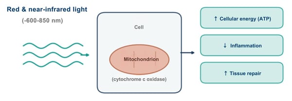
Figure 1. How PBM works. Red and near-infrared light penetrate tissue and are absorbed by mitochondrial cytochrome c oxidase, raising cellular energy and triggering downstream repair and anti-inflammatory effects.
In November 2024, LumiThera’s Valeda system became the first FDA-authorized treatment of any kind shown to improve vision in dry age-related macular degeneration, a leading cause of central vision loss for which no prior therapy had been approved.
The pivotal LIGHTSITE III trial randomized 100 subjects, met its primary visualacuity endpoint with multiwavelength light at 590, 660, and 850 nm, and a 2025 extension reported that the gains held with continued treatment. (al., 2026, pp. 1-9) For a field long dismissed as soft, an FDA authorization backed by a shamcontrolled trial is a different order of evidence.
Another clear proof that PBM works lives in oncology. Oral mucositis, the painful breakdown of the mouth's lining, strikes most patients on certain chemotherapy and radiation regimens. (Atwiine et al., 2024, pp. 354-364) It also responds to red and near-infrared light. The evidence is strong enough that PBM receives a full recommendation in the 2020 MASCC/ISOO clinical guidelines for preventing it in patients undergoing head-and-neck radiotherapy and stem-cell transplantation. (Elad et al., 2020, pp. 4423-4431)
MuReva Phototherapy, spun out of Lumitex in 2018, provides a concrete example of this clinical translation. Its MuReva OM system uses a soft, flexible intraoral mouthpiece and integrated lightguide to deliver 660 nm light simultaneously across tissues susceptible to oral mucositis. In November 2025, FDA granted the prescription device De Novo authorization, establishing a new Class II category for intraoral phototherapy devices.
MuReva’s progress illustrates a broader truth about PBM: successful clinical adoption depends not only on the biological effects of light, but also on controlled dosing, practical delivery and a form factor that works for patients and care teams.
This places therapeutic light among the standard supportive-care tools in cancer treatment, and newer work is moving it from the clinic into self-applied, home-use devices.
Overall, PBM has a broad, active body of work in wound healing, accelerating repair in diabetic and other slow-healing wounds. (Torkaman et al., 2024, pp. 275-284) It has its largest real-world footprint in pain and musculoskeletal care, the home of most cleared devices and the most familiar face of therapeutic light, though the quality of the evidence varies widely by indication. ((NICE), n.d.)
At the frontier sits transcranial PBM, delivering light through the skull to influence the brain, among the most intriguing directions in the field and among the least settled (it was the subject of a prior edition of this report). (Taylor et al., 2024, pp. 822-827)
The current literature and recent developments on PBM are real footholds. So why does a therapy with guideline backing, an FDA authorization, and 60 years of research behind it not see more use?
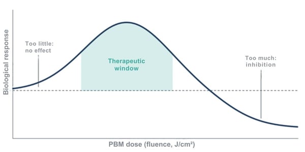
Figure 2. The biphasic or Arndt-Schulz dose response. Too little light produces no effect, while an intermediate therapeutic window yields benefit. Too much suppresses the response, making the correct dose a narrow target.
No consensus dosing: PBM follows a biphasic dose-response, often described by the Arndt-Schulz curve. Too little light does nothing, and too much can actively inhibit the response you wanted. That makes the right dose a narrow target. There is still no consensus framework for defining that therapeutic window across different tissues, depths, and diseases.
Breadth is a liability: PBM appears to help to some degree with an enormous range of conditions, and that scope actually works against it. The field itself identifies the sheer breadth of PBM applications as a credibility barrier. A therapy that seems to treat everything reads, to a skeptical clinician, as one that proves nothing.
Cleared, not approved: Most PBM devices reach the U.S. market through general clearances for broad indications, such as a temporary increase in local blood circulation or relief of minor muscle and joint pain, rather than authorization for a specific disease. The dry AMD and OM authorizations stand out precisely because they are the exceptions.
Reimbursement lags evidence: Even PBM’s biggest clinical and regulatory wins are still works in progress. Retinal PBM received Category III CPT code 0936T, effective January 1, 2025. MuReva’s intraoral PBM treatment for oral mucositis followed with Category III CPT code 1011T, effective January 1, 2026. These emerging-technology codes give providers a defined way to report the procedures and allow utilization data to accumulate, but they do not guarantee payer coverage or payment. Until payers establish consistent reimbursement, adoption can remain slow regardless of the strength of the clinical evidence.
None of these obstacles are permanent, and none are optical. Standardized dosimetry would make trials reproducible for regulators and payers. The dry AMD authorization gives the next developer a template to follow, one disease at a time. And as clinical grade systems separate from the wellness-gadget market, the credibility drag eases.
The real constraints for PBM were never optical, but rather the absence of standardized dosing, the gap between broad clearance and disease-specific proof, the slow arrival of reimbursement, and the credibility drag of a market full of unproven gadgets.
With the first meaningful authorization on the board and a visible pathway behind it, the next chapter for PBM is about discipline rather than discovery, turning a 60-year promise into routine clinical practice.
The science has been ready for a while. The pieces are starting to line up. And the field is finally catching up.
With the first meaningful authorization on the board and a visible pathway behind it, the next chapter for PBM is about discipline rather than discovery, turning a 60-year promise into routine clinical practice.


Insulin is one of the most important drugs in medicine, and also one of the bluntest. Once injected, it works on its own schedule, not the body’s. Give a little too much, and a patient can slide into hypoglycemia, a drop in blood sugar that ranges from unpleasant to life-threatening.
For a hormone whose entire job is precise regulation—and for the hundreds of millions of people who depend on it—insulin’s lack of fine control is a real limitation.
Now picture a different approach: cells inside the body that produce insulin only when instructed, in measured amounts, on a timescale of minutes, with the onswitch being nothing more than a pulse of light.
That is the premise behind optogenetics. And for diabetes care, the cutting-edge frontier of medical lighting has already moved from idea to measurable results in animal trials.
Now picture a different approach: cells inside the body that produce insulin only when instructed, in measured amounts, on a timescale of minutes, with the onswitch being nothing more than a pulse of light.
Optogenetics is the practice of engineering cells to respond to light, and the basic process is more approachable than the name suggests. A delivery vehicle, often a harmless virus, carries a set of genetic instructions into a target cell. The cell reads those instructions and builds a light-sensitive protein it would not normally produce. When a specific wavelength of light reaches that protein, it flips the biological switch on or off, causing the cell to respond in a specific, pre-engineered way. In this case, the response is increased insulin production.
It’s worth pausing here because this sounds a great deal like Photobiomodulation, the light therapy covered earlier in this report. Photobiomodulation works with the light-sensitive molecules a cell already has, catalyzing existing biology. Optogenetics installs a brand-new, synthetic switch that the cell never had before. One influences, while the other controls.
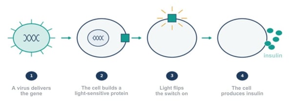
Figure 1. How optogenetics works. A gene-delivery vehicle carries instructions into a cell, which builds a light-sensitive protein. A pulse of light flips the switch, and the cell then produces insulin.
For optogeneticists, precision control is the point. Because the switch is engineered, researchers can dictate not just whether a cell acts but exactly when and how much, with a precision in space and time that ordinary drugs cannot match.
Unsurprisingly, optogenetic approaches are under study for eye disease, cancer, and metabolic disorders, but diabetes is where recent momentum is most evident.
A recent generation of light-switchable systems has demonstrated fast, reversible control of insulin in living animals, providing a different order of evidence for a promising mechanism on paper.
REDMAP uses red light around 660 nanometers to activate insulin gene expression. In diabetic mice and rats, light exposure could turn the switch on and lower blood glucose. Red light matters for a practical reason: it penetrates tissue more deeply than shorter wavelengths, so a red switch is an ideal candidate for use beneath the skin.
A second approach uses blue light to drive rapid insulin secretion. While the redlight switch works at a slower level of gene expression, this one acts within minutes. In one study of diabetic mice, insulin levels rose within about 15 minutes of blue-light exposure, and blood glucose returned to a non-diabetic range within an hour. (Chen et al., 2024)
Taken together, the two illustrate an important point. Different wavelengths can be matched to different jobs (e.g., slow and deep vs. fast and shallow), suggesting therapies that could one day be tuned to a patient’s moment-to-moment needs.
For now, these are just animal results. None of this has been tested in humans for the treatment of diabetes. But moving from interesting biology to measurable glucose control on a clinical timescale is exactly the kind of step that propels a viable medical technology toward clinical use.
Several problems remain to be solved before practical optogenetic applications will be ready for human trials. Most of them are not about whether the biology works:
Light spectrum: Visible light, especially blue light, does not travel far through tissue. Reaching cells deep in the body with enough light to flip a switch is the field’s central physical obstacle.
Delivery mechanism: Getting the synthetic switch into the right cells, and only the right cells, depends on gene-delivery tools that are still being made safer, both the viral kind and newer non-viral approaches.
Cell engineering: The engineered cells that carry the switch, sometimes called chassis cells, have to remain safe and stable in the body over long periods, which is a high bar for any cell-based therapy.
The optogenetic therapies that have reached clinical trials so far treat neurodegenerative eye diseases such as retinitis pigmentosa, and for a telling reason. In one widely reported case, a patient who had been blind for decades regained partial vision after receiving a light-sensitive protein in the retina paired with a set of light-projecting goggles.
Zooming out, the eye is the ideal proving ground because the retina sits just behind the lens, already built to receive light, which sidesteps the penetration problem entirely. Diabetes, which occurs deeper in the body, is a harder target.
Almost every remaining obstacle to the adoption of optogenetics ultimately comes down to the same question: how do you get the right light to the right cells? The next wave of work is increasingly an engineering problem rather than a biological one.
Currently, two design approaches aim squarely at the depth problem:
Designing light-responsive proteins: These would respond to near-infrared light, which reaches deeper into tissue than visible wavelengths. Upconversion nanoparticles absorb deep-penetrating near-infrared light and re-emit it as visible light that the switch needs, carrying a usable signal where visible light cannot reach.
Embedded Light: Stop sending light through tissue at all. Implantable, wirelessly powered light sources can be placed exactly where the engineered cells live, completely solving the penetration problem.
For diabetes, the destination these threads point toward is a closed loop. A wearable sensor measures blood glucose, a controller determines a dose, and a light source signals the engineered cells to release insulin in real time, continuously and noninvasively.
Early versions of exactly this loop have already been demonstrated in diabetic mice, pairing a glucose-sensing component with implanted, light-controlled cells. It would turn diabetes management from a series of manual corrections into something closer to automatic treatment.
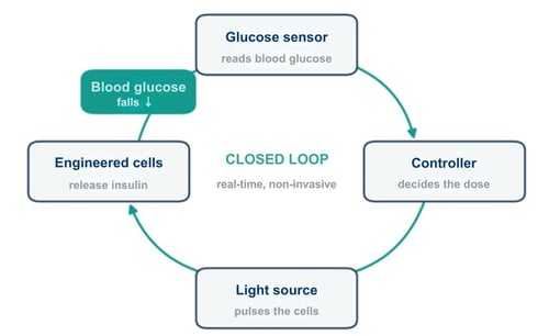
Figure 2. The closed-loop goal for diabetes. A wearable sensor reads glucose levels, a controller sets the dose, a light source signals the engineered cells, and the insulin they release lowers glucose levels.
None of this means insulin injections are going away anytime soon. For diabetes, optogenetics is still early and still pre-human, and the road to the clinic runs through real questions about safety, delivery, and depth. Overpromising would do the field no favors.
But the trajectory here is hard to miss. Medicine has spent a century getting better at identifying and responding to chemical signals in broad, slow strokes. Optogenetics points to something different: control that is precise, reversible, and timed to the second.
Medicine has spent a century getting better at identifying and responding to chemical signals in broad, slow strokes. Optogenetics points to something different: control that is precise, reversible, and timed to the second.