

%20(2240%20%C3%97%201120%20px).png?width=2240&name=Surgical%20Lighting%20(Blog%20Banner)%20(2240%20%C3%97%201120%20px).png)
Surgical lighting ergonomics is a hot topic in the surgical world. Overhead lights can have limited positioning and may not provide adequate visibility in deep tissue or minimally invasive surgery. Headlamps are popular, but can be extremely cumbersome and can add strain to the neck during long surgeries, especially with additional COVID protective equipment.
One of the biggest and most concerning ergonomic issues caused by surgical lighting can be that unmodulated high-intensity light can result in eye strain and fatigue.
In this article, we will focus on surgical lighting ergonomics related to eye strain and visibility during procedures and new trends that help improve these issues.
Surgical Lighting Ergonomics
Studies suggest that unmodulated high-intensity light can result in eye strain and fatigue. A survey revealed that 25% of surgeons report that eye strain is an occupational health hazard (3). According to the American Academy of Ophthalmology, one of the main causes of eye strain (asthenopia) is excessive or improper illumination. Eye strain is also aggravated by rapid eye movements required for detail work and is seen more often in minimally invasive surgery (11).
Symptoms of eye strain can include pain in & around the eyes, headache, fatigue, vertigo, facial muscle tics, or migraine. Mitigation of can involve using correcting underlying issues such as corrective lenses, adjusting lighting, and taking breaks for eye rest in cases of prolonged detail work (10). Since it’s not practical to take visual breaks during surgery, lighting levels should be optimized to reduce eye strain and fatigue. In addition, optimal light spectra can be used to improve visualization, making intraoperative decisions easier and reducing surgery time.
Improved Visualization During Procedures
Different CRI and wavelengths emitted from light sources reveal tissues differently. The way tissues appear visually is an important aspect of assessment and diagnostics during procedures. Keep reading to find out how LEDs are being used to detect cancer and better visualize tissues in surgical procedures.
Surgical Lighting Trend—The Switch to LED Light Sources
Acceptance of LED Sources in Medical Lighting
While halogen lights are considered the “gold standard” in surgical lighting, LED light sources are gaining popularity (6). Halogen sources are widely available, while LED technology has gradually improved over the last several years. LEDs are often preferred because they require less energy to operate, reducing heat output.
Traditional light sources such as halogen bulbs represent a visual light spectrum most like natural daylight, allowing for optimal visualization during procedures. In contrast, LEDs can be heavily weighted to the blue end of the visible light spectrum. This often causes a “washed-out” appearance. If exposed for too long, blue-heavy light sources also have negative effects on eye tissues and the brain(9).
As a result of these issues, acceptance of LED light sources has been slow. Fortunately, LED technology has improved spectral output, resulting in more accurate color rendering. This progress allows us to benefit from both the high-fidelity color of traditional sources and the efficiency of LEDs.
Color Rendering
What is Color Rendering?
The most common way to assess a light source’s color output is the Color Rendering Index or CRI. This index was developed by the International Commission on Illumination (CIE) starting in the 1930s and has undergone several changes to capture the way different sources display color more accurately. The CRI measures colorimetric information, or how the human eye perceives color. The CRI is a complex calculation using the hue and saturation of a color swatch set and the source's correlated color scale (CCT).
How does Color Rendering Work?
Natural, bright sunlight is given a score of 100 and is the basis of comparison for all other lighting. Typically, incandescent and halogen bulbs score high on the CRI scale. Some LEDs do not score well on the CRI scale and tend to lend themselves toward color desaturation. With recent advances, the CRI of LEDs has improved, as some manufacturers claim a CRI higher than 90.
The images below show examples of spectral power distribution (SPD) curves. Notice the spectral distribution difference between standard tungsten, filtered daylight, and a LED-based source.
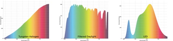
Source: National Gallery: Spectral Power Distribution (SPD) Curves by Joseph Padfield licensed under (CC BY-NC 4.0)
These differences result in the different color renderings of the visual target.
Spectral power distribution is specific to each light source and affects how color is rendered. The distribution shows the wavelength and intensity of light in the visible spectrum. Sunlight has an even distribution, making it the standard of comparison for artificial light sources. Interestingly, the Academy of Motion Pictures completes detailed research on the spectral output of LED arrays and the effect on color rendering.
You can look at their article here: Solid State Lighting Report
There are several ways to define color rendering. The Correlated Color Scale and the Color Quality System are two well-defined ways we can understand the appearance of colors under different light sources.
Correlated Color Scale
Correlated Color Temperature Scale (CCT) is a scale in degrees Kelvin. It describes the color appearance of the light source, which affects the perception of colors in the environment. The scale uses a black body radiator to determine a lighting temperature.
A black body radiator is a theoretical concept that describes an ideal source of thermal radiation that does not reflect any heat. As the black body increases in temperature, it emits visible light. As it continues to heat, its color changes from black to red to yellow, white, and blue. A specific temperature causes the black body to turn a specific color, and that temperature/color is expressed in degrees, Kelvin. “Cooler” color temperatures, like white and blue, are on the higher end of the temperature scale.
The black line (or the Planckian locus) on the image below shows the colors a black body radiator emits as it is heated (right to left). The x and y coordinates indicate the chromaticity of the color. Chromaticity is a specification of the hue and saturation of a color.
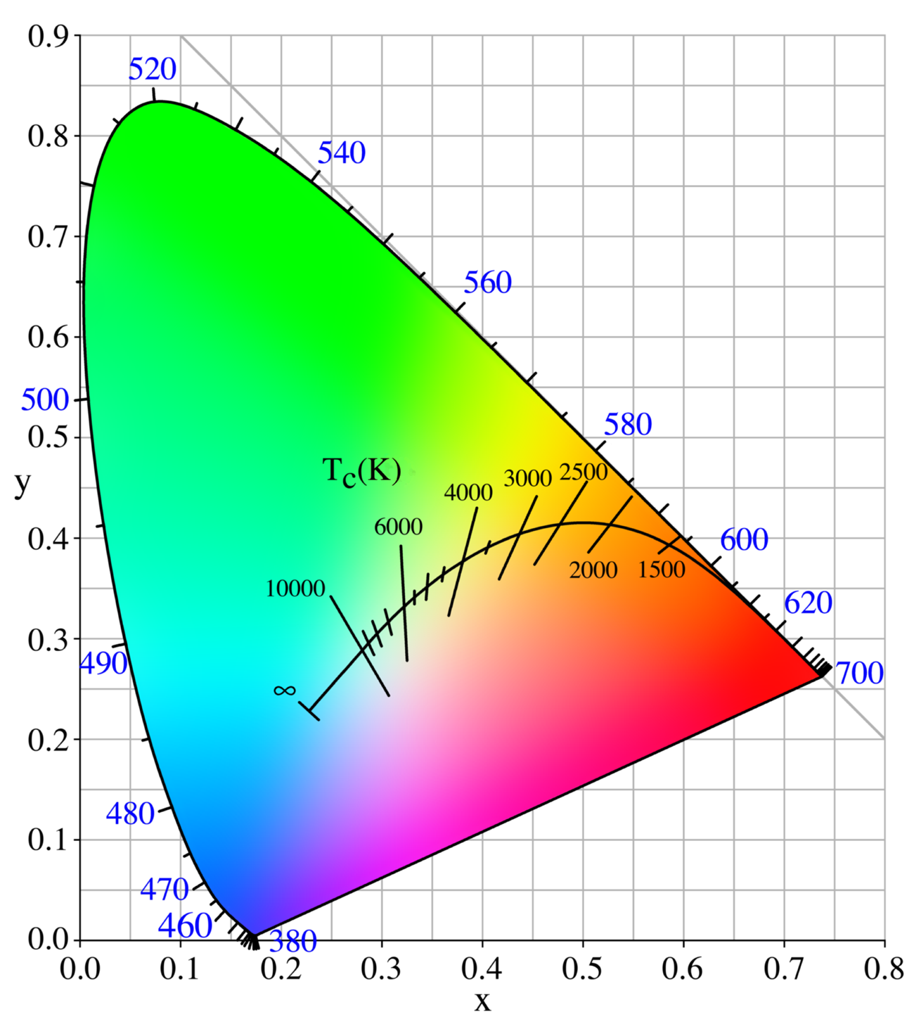
Source: Wikimedia Commons. Planckian locus. en: User: PAR, Public domain, via Wikimedia Commons.
It is common to see these color temperatures listed as a specification on LED bulbs in addition to a description such as “Cool White” for a 7000K bulb or “Warm White” for a 2000-3000K bulb.
How Colors Look Under the CCT Scale
Below are test color sample swatches as they appear under daylight. The first 8 are the original CRI color swatches; the remainder has been added to evaluate light's ability to render color more accurately as LEDs have increased in popularity. These swatches are standardized to evaluate the color rendering of a light source. If the color swatches closely match those under daylight, the light source is said to have a high CRI. If the colors appear washed out or have a different hue, the CRI is poor.
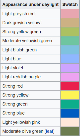
Source: Wikipedia. Color rendering index.
Despite all these factors that determine color rendering index, the CCT scale is an incomplete system. One of the main issues is that the representative color swatches don’t represent the full visible color spectrum. Notably, red is greatly under-represented in the color sampling swatches.
Color Quality System
NIST has developed a new color quality rating system in conjunction with the lighting industry and the International Commission on Illumination (CIE). The Color Quality System (CQS) uses a larger number of color samples that span the entire visible light spectrum at equal intervals. The CQS score is also calculated differently to ensure that the rating is not improperly skewed if one color sample is shifted more than others.
CQS also incorporates other color quality factors like contrast. While this rating system gives a complete description of object color appearance, CRI has been used in the industry for over 40 years and is widely used by manufacturers.
Importance of Color Rendering in Medical Applications
Why is color rendering so important?
LED technology is progressing to the point that tuning spectrum distribution enhances contrast in medical lighting. LEDs have been used to detect oral cancer and improve texture visibility (7).
Recent research projects have shown that enhancing certain spectra can improve the visibility of tissues. For example, the 450-550 nm range enhances the visibility of blood vessels surrounded by muscle tissue (7). By utilizing LEDs with specific spectral outputs, it can be easier to identify lesions and target tissues during a procedure (7,8).
By blending white light LEDs with specific spectra LEDs, an ideal mixture of medical lighting can be achieved while minimizing eye strain. These tunable sources are currently available, but more work is being done on wide acceptance and efficacy.
In addition to color rendering for viewing, work is being done to make tunable operating room lighting to improve eye comfort for patients and staff. A Danish company has been making ergonomic lighting that improves surgeon and staff viewing for LED laparoscopy screens. These systems also reduced glare and improved radiograph reading and venipuncture (5).
Choosing a light source that accurately illuminates the environment results in a better viewing experience. From reducing eye fatigue to improved visual accuracy, a high color rendering index makes the light source a part of the background—something that the viewer doesn’t even have to think about. Our goal at Lumitex is to provide a visual and lighting experience that appears natural and intuitive. We measure our success by how little your end-user needs to think about lighting or eye strain.
FAQs About Surgical Lighting Ergonomics
Why is surgical lighting ergonomics important?
Surgical lighting ergonomics helps reduce eye strain, fatigue and discomfort during long procedures. Proper lighting placement and balanced illumination improve visibility and support surgeon performance.
How do LED surgical lights improve operating room visibility?
LED surgical lights improve visibility by providing better energy efficiency, lower heat output and improved color rendering. Modern LED systems also help reduce glare and visual fatigue during procedures.
What is color rendering in surgical lighting?
Color rendering describes how accurately a light source displays colors compared to natural daylight. High color rendering helps surgeons distinguish tissues, blood vessels and lesions more clearly during procedures.
Why can high-intensity surgical lighting cause eye strain?
Unmodulated high-intensity lighting can force the eyes to constantly adapt to glare and brightness differences. This can lead to headaches, fatigue and reduced visual comfort during surgery.
How does spectral output affect medical lighting?
Spectral output affects how tissues appear under surgical lighting. Specific wavelengths can improve contrast and help clinicians better identify anatomical structures during procedures.
Why are LEDs replacing halogen surgical lights?
LED lighting systems are becoming more popular because they consume less power, generate less heat and now provide improved color rendering compared to older lighting technologies.

11. Stucky, Chee-Chee H. et al. “Surgeon symptoms, strain, and selections: Systematic review and meta-analysis of surgical ergonomics”. Annals of Medicine & Surgery. 27 (2018) 1-8

Comments